The Volume of Subscapularis Muscle Remains Unaffected by Supraspinatus Tendon Tears: Three-dimensionally Reconstructed Magnetic Resonance Imaging Analysis
Article information

Abstract
Background
This study aimed to compare the subscapularis muscle volume between the intact groups (group I) and supraspinatus tendon tear groups (group T) based on the sex and three different age groups.
Methods
Subjects with a group I and subjects with group T without any other lesions were retrospectively evaluated from among patients who received a magnetic resonance imaging (MRI) scan between January 2011 and December 2013. The MRI scans were studied by a consultant radiologist. The subscapularis muscle volume was compared according to the age and sex; the age groups were categorized as patients in their 40s, 50s, and 60s. The volume of subscapularis muscle was measured by three-dimensional reconstructed images acquired through the axial section of 1.5T MRI.
Results
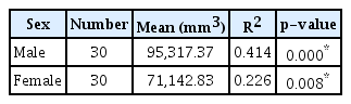
No statistically significant differences were observed between subscapularis muscle volume of the group I and group T, except for male patients in their 50s (group I: 100,650 mm3 vs. group T: 106,488 mm3) and 60s (group I: 76,347 mm3 vs. group T: 99,549 mm3) (p<0.05). Males had a larger mean volume of subscapularis muscle than females, and the subscapularis muscle volume decreased in a linear manner with increasing age.
Conclusions
Decrease in subscapularis muscle volume was observed with increasing age, and the impact of supraspinatus tear on subscapularis muscle volume is age and sex dependent.
Introduction
Due to the inherent interaction with each other, the four rotator cuff muscles serve as the main dynamic stabilizers of the humeral head [1-3]. The antagonist subscapularis and infraspinatus/teres minor muscles build a force couple that centers the humeral head in an anteroposterior direction [4,5]. Any asymmetric tonus of the agonists results in altered glenohumeral shear force leading to humeral head displacement from its center of rotation in the glenoid cavity, thereby resulting in the other antagonist muscle weakness and atrophy [5,6]. Also, changes in muscle size and joint strength are known to occur throughout the adult lifespan. Muscle volume, especially of the shoulder, decreases with age. The linear relationship, however, between muscle volume and age remains uncertain [7-9].
The subscapularis muscle is the largest and most powerful muscle among the rotator cuff muscles; it is also named the “forgotten tendon” as it rarely tears. This muscle plays a key role as an anterior structure of the transverse force couple along with the infraspinatus and teres minor muscles, which are posterior to the shoulder joint [10,11]. Among the posterior structures of the transverse force couple, the importance of the infraspinatus and teres minor muscles are widely recognized; however, significance of the supraspinatus muscle as the posterior structure of the shoulder joint remains unclear.
Recently, magnetic resonance imaging (MRI) has been widely applied to evaluate the shoulder joint, and has been highly useful in understanding anatomical structures [12-15]. Several methods have been introduced to measure the volume of the rotator cuff muscles. Tingart et al. [16] separated each muscle of the rotator cuff in cadaver experiments and then measured the amount of overflowing water when each muscle was soaked in water. Juul-Kristensen et al. [17] evaluated the muscle volume in 20 patients using MRI. According to the data collected by Juul-Kristensen et al. [17] and Tingart et al. [16], no differences were observed between the volume of muscle obtained by soaking in water in the cadaver experiments and the volume of muscle obtained by three-dimensional (3D) reconstruction with MRI. Currently, rotator cuff atrophy is assessed using a multi-layered approach involving these methods.
As a posterior structure of the shoulder, the supraspinatus tear affects the posterior shoulder force couple, and could also affect the anterior shoulder muscle volume (subscapularis). This retrospective study was undertaken to evaluate if the supraspinatus tendon tear affects the subscapularis muscle volume. We compared the subscapularis muscle volume between groups, with and without supraspinatus tendon tear, by considering sex and age.
Methods
Subject Enrollment
Between January 2011 to December 2013, 1,592 patients underwent MRI due to shoulder pain. We analyzed MRI readings of 1,218 patients in their 40s to 60s; 374 patients were excluded since they did not meet the age requirement. The results were studied by a musculoskeletal radiologist having an experience of more than 30 years. Among the 1,218 patients enrolled, 990 patients were excluded due to abnormal MRI findings, including supraspinatus tendon tear. Of the 228 patients finally enrolled for evaluation, 96 patients had only supraspinatus tendon tear (group T) and 132 patients showed no abnormality (intact) in the MRI (group I). Considering the age, 60 subjects were randomly selected from both groups, where 20 each belonged to the 40s, 50s, and 60s age groups (Fig. 1). Subjects were randomly selected from each subgroup using random sampling in Excel (Microsoft, Redmond, WA, USA).
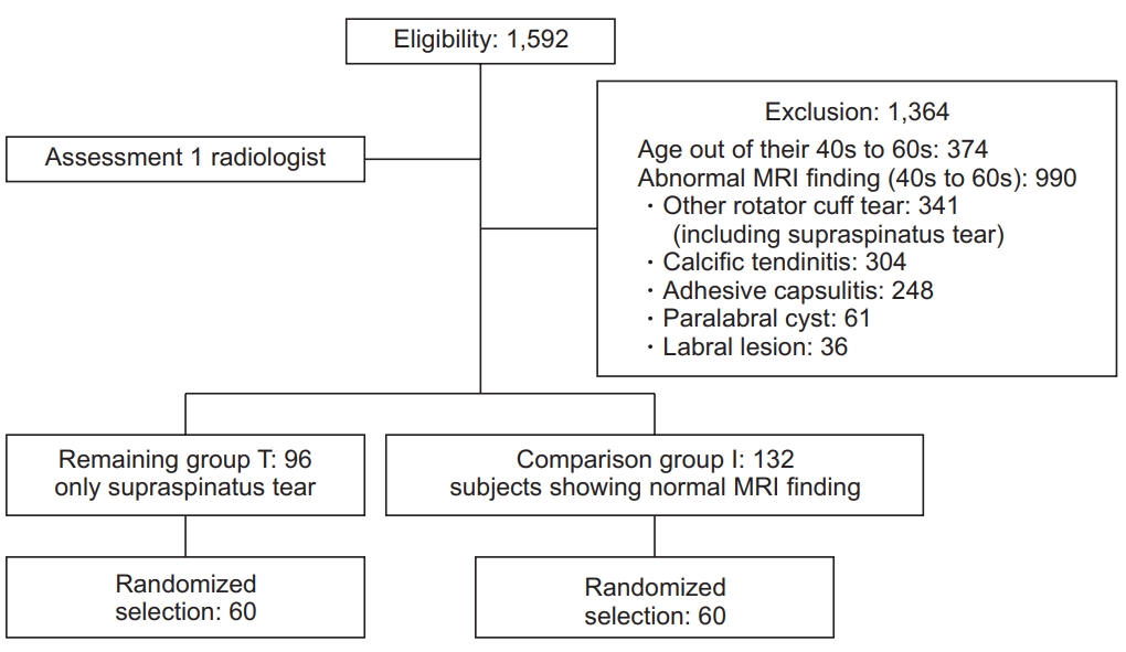
Flow chart of subject selection. Group I showed no abnormality (intact) and group T had only supraspinatus tendon tear in the magnetic resonance imaging.
MRI: magnetic resonance imaging.
Demographics
A total of 120 patients were evaluated, including 60 shoulder joints in female patients and 60 shoulder joints in male patients, with an overall mean age of 54.3 ± 9.29 years (range, 40–69 years). Totally, 60 participants had a normal supraspinatus tendon (group I: age range, 40–69 years; mean, 55.1 ± 9.47 years), and 60 patients had a supraspinatus tendon tear (group T: age range, 40–69 years; mean, 53.6 ± 10.32 years). The three categorized age groups consisted of 20 patients each.
Three-dimensional Reconstruction and Volume Measurement
We conducted a quantitative assessment of the subscapularis muscle volume using 3D reconstruction of MRIs in patients included in the group T and group I. The MRIs used in this study were obtained through an Avanto 1.5-T MRI (Siemens, Erlangen, Germany). The slice size of the images was 3.0 mm.
Briefly, muscle contours were assigned to every transverse slide of the MRI by using the ‘‘Livewire’’ option on the MIMIC 10.01 (for Intel x86 Platform III+; Materialise NV, Leuven, Belgium) medical imaging software. Volume masks were calculated from the assigned contours on the transverse slides. The subscapularis muscle was outlined on each scan, proceeding from the medial border of the scapular to the lesser tuberosity, which belongs to the subscapularis tendon. As the outline was hand-drawn in the MIMIC program, accuracy was low. To increase the accuracy, 3 orthopedic surgeons participated in taking measurements (Fig. 2, 3). Interobserver reliability was evaluated using intraclass correlation coefficients (ICCs) introduced by Shrout and Fleiss [18] Interobserver reliability was relatively high, with an ICCs of 0.836.
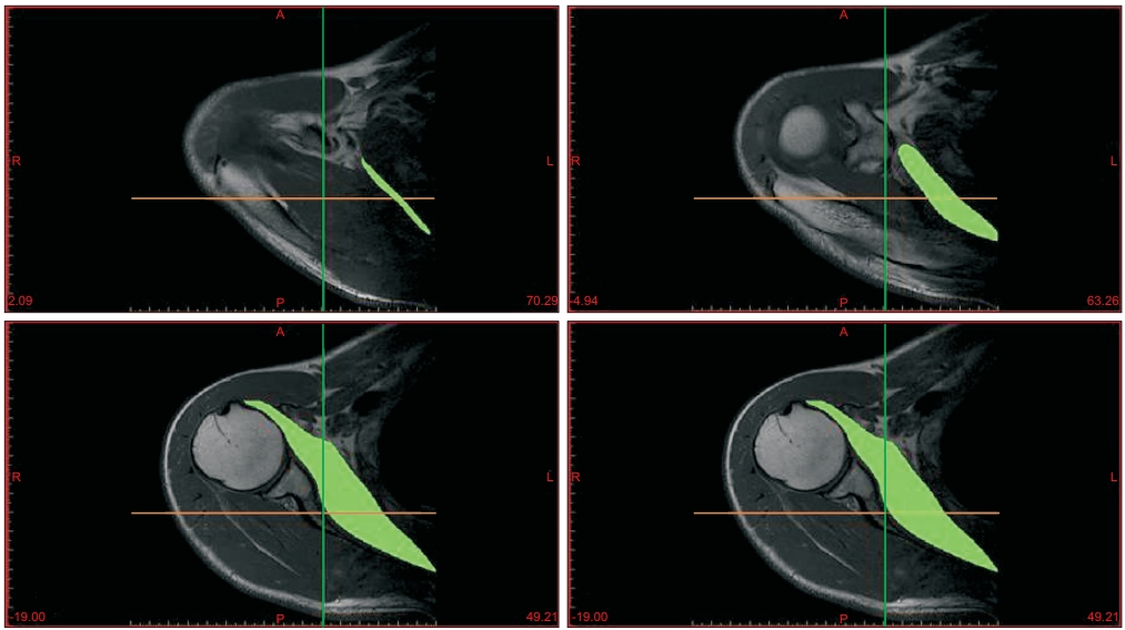
Green label: Schematic surface of the subscapularis muscle border in a transverse section; Green colored schematic surface of the subscapularis muscle on transverse section can be seen here; Drawing the outline of the subscapularis muscle in a transverse section.

Three-dimensional (3D) reconstruction image of subscapularis muscle. (A) 3D reconstruction of the image of the subscapularis muscle is presented. Subscapularis 3D reconstruction image; (A) is the anterior aspect of the 3D reconstruction image of subscapularis muscle, (B) is the posterior aspect, (C) is the anteroinferior aspect, (D) is the anterosuperior aspect.
Statistical Analysis
All data were statistically analyzed using the IBM SPSS ver. 21.0 software (IBM Corp., Armonk, NY, USA). Continuous variables between the two groups was compared by the t-test (Table 1), and linear regression was used to analyze the correlation between age and subscapularis muscle volume according to sex (Table 2-4). Statistical significance was accepted at p<0.05.

Age Specific Subscapularis Muscle Volume and t-test of Supraspinatus Tendon Tear Patients and Normal Participants
Linear Regression Analysis of the Volume of Subscapularis Muscle in Normal Participants (group I)

Linear Regression Analysis of the Volume of Subscapularis Muscle in Patients with SST (group T)
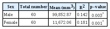
Linear Regression Analysis of the Total Volume of Subscapularis Muscle (group I and group T)
Results
No significant differences were detected between the groups with respect group I and group T among participants in their 40s (p>0.05). However, a significant difference was observed between both groups when considering male in the 50s and 60s (p<0.05). Subscapularis muscle volume loss tended to increase with age in the group I, as indicated by linear regression analysis (Fig. 4, 5). However, this linear decrease was not observed in the group T (Table 2, 3).
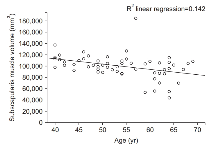
Linear regression analysis of the age and subscapularis muscle volume in males. The average volume of subscapularis muscle is decreased in males, with aging.

Linear regression analysis of age and subscapularis muscle volume in females. The average volume of subscapularis muscle is decreased in females, with aging.
Discussion
The primary goal of this study was to explore the relationship between the subscapularis muscle volume and supraspinatus tendon tear. Results of this study indicate no significant relationship between supraspinatus tendon tear and volume of subscapularis muscle in females. However, a higher subscapularis muscle volume was observed in group T than group I of males in their 50s and 60s.
We hypothesized that the posterior section of the supraspinatus interacts with the subscapularis muscle as a posterior structure which contributes to some degree of anterior-posterior stability. Furthermore, the supraspinatus tendon tear affects the volume of the subscapularis muscle loss, thereby leading to a tear [19].
According to Bergin et al. [19], subscapularis tendon abnormality is related to chronicity of supraspinatus tendon tears. In patients with supraspinatus tear, the dynamic anterior instability increases due to the elevated posterior shoulder force couple weakness, resulting in sub-coracoid impingement and consequently a subscapularis tendon tear [19]. MRI scans reveal the existence of bone marrow edema of lesser tuberosity; however, the study of bone marrow edema was not included in the current study.
Contrary to our hypothesis, our study revealed different outcomes. We initially hypothesized that supraspinatus tendon tear affects the reduction of the subscapularis muscle volume. However, we did not account for the degree of supraspinatus tendon tear size, tear thickness, torn tendon retraction degree, occupation, activities, and body skeletal mass [20]. Hence, our data revealed no significant relationship between supraspinatus tendon tear and volume of subscapularis muscle in the group T of males in their 40s. Since the supraspinatus tendon tear might be small in the 40s with a shorter duration of tear, its effect on the volume of the subscapularis muscle may be negligible. Furthermore, in the group comprising males in their 50s and 60s, group T had higher subscapularis muscle volume than group I. We also considered that males included in the group T had a higher level of labor intensity or sports activities, and these factors might affect in increasing the volume of subscapularis muscle before onset of supraspinatus tear. However, we also considered that this result could be due to the small number of samples, thereby resulting in selection bias which plays a role in enhancing the subscapularis muscle volume in group T than in group I. Furthermore, our data showed that the subscapularis muscle volume in group T of female was lower than in group I, but was statistically not significant. We believe this could be that most female are housewives or working with lower labor intensity jobs. When considering sex of the female, the subscapularis muscle volume seemed more affected by the supraspinatus tendon tear than by life activities.
In this study, we found that although the subscapularis muscle volume reduces with aging, it was not related to cuff tear. This finding is supported by previous muscle studies that demonstrate that the aging process results in a loss of muscle mass, with subsequent replacement by fat and connective tissue [21,22]. Regardless of the sex, reduced muscle volume and aging were correlated for subjects in their 40s, 50s, and 60s. With the onset of advancing age, muscle tissue is gradually lost, resulting in diminished mass and strength; this condition is referred to as sarcopenia [21,23]. One study presented that patients with cuff tears have sarcopenia, and patients with large to massive tears have a significantly inferior sarcopenic index than those with small to medium tears [24]. They measured the whole-body muscle mass as per the Janssen [25] method of bioelectrical impedance analysis equation, which is different from the analysis used in our current paper, in that we only calculated the subscapularis muscle [24]. Another paper explains that rotator cuff atrophy is related to increasing age and not to tear severity. For patients without rotator cuff tears, the prevalence of fatty infiltration and atrophy increased with aging [26]. Further evaluation is required to determine the pathophysiology and progression of muscle atrophy in rotator cuff muscles.
Although cadaver studies provide a detailed understanding of the effects of supraspinatus tendon tear on the subscapularis muscle atrophy or tendon tear, it is necessary to achieve meaningful results. In this study, we measured the muscle volume of the rotator cuff (such as subscapularis muscle) through MRI. Since only the muscle part is measured (except for the surrounding fatty tissue), the accuracy is relatively high [27]. Considering various other factors such as tear size, jobs, sports activities, height, and weight that influence the muscle mass will yield more meaningful results.
This study has some limitations. Most notably, being retrospective in design, we are unable to make any causal claims explaining the relationship between supraspinatus tear and subscapularis muscle volume. Second, this study has a small sample size and does not exclude factors that might affect muscle volume, such as height, weight, occupation, and sports activities. Third, we did not account duration of the disease and size (i.e., full-thickness tear or partial thickness) of the supraspinatus tendon tear. Fourth, muscle volume measurements using the MIMIC program did not consider fatty infiltration in the muscles, hence the measured volume value does not reflect the intrinsic atrophy of the muscles accurately.
Conclusion
Analysis applying 3D reconstruction by MRI reveals decreasing subscapularis muscle volume with aging, regardless of the sex. The supraspinatus tear was seen to affect the subscapularis muscle volume in a certain group of age and sex.
Notes
Research Ethics
IRB approval: Chosun University Hospital (No. CHOSUN 2015-11-003-002).
Conflict of interest
None.
Financial support
None.