Cubital tunnel syndrome associated with previous ganglion cyst excision in the elbow: a case report
Article information

Abstract
Cubital tunnel syndrome refers to compression neuropathy caused by pressure on the ulnar nerve pathway around the elbow. A 63-year-old male patient visited the clinic complaining of decreased sensation and weakness in his left ring finger and little finger, stating that the symptoms first began 6 months prior. He had undergone surgery to remove a ganglion cyst from his left elbow joint about 5 years prior in Mongolia. Magnetic resonance imaging revealed a cystic mass located at the previous surgical site, which was compressing the ulnar nerve within the cubital tunnel. Ulnar nerve decompression and anterior transposition were performed, and the cystic mass was excised. Upon pathological examination, the mass was diagnosed as a ganglion cyst. The patient’s symptoms including sensory dysfunction and weakness improved over the 1-year follow-up period. This report describes a rare case of ganglion cyst recurrence compressing the ulnar nerve in the cubital tunnel after previous ganglion cyst excision.
Level of evidence
V.
Cubital tunnel syndrome (CuTS) is the most common form of ulnar nerve entrapment and the second most common form of compressive peripheral neuropathy [1]. It generally occurs due to the compression of structures such as the arcade of Struthers, medial intermuscular septum, Osborne’s ligament, deep fascia of the flexor carpi ulnaris (FCU), fibrous arch between the humeral and ulnar portions of the FCU, and deep flexor-pronator aponeurosis [1]. Other common causes include osteoarthritis, direct trauma to the elbow joint, fracture of the lateral humeral condyle, dislocation of the elbow joint, and deformities of the cubitus valgus and varus. It can also be caused by synovial hyperplasia, soft tissue abnormalities such as tumors, and secondary nerve compression from cysts such as ganglion cysts [1]. Ganglion cysts that cause CuTS can be classified as intraneural or extraneural ganglion cysts. Complete excision of extraneural ganglion cysts is required, whereas the decompression of hypertrophic nerves and removal of the articular branch of the cyst are recommended for intraneural ganglion cysts, which are rare [2]. While ganglion cysts are often asymptomatic, they may cause pain or weakness due to the compression of nerves and blood vessels and can also lead to esthetic concerns, which can be indications for surgery [3,4].
We report the rare case of a patient who experienced an improvement in numbness and weakness following surgical intervention for the recurrence of a ganglion cyst at the site of previous ganglion removal surgery, and a corresponding literature review.
CASE REPORT
This study was approved by the Institutional Review Board of Sungae Hospital (No. SA2022-05), and informed consent was obtained from the patient. A 63-year-old Mongolian man was admitted with complaints of altered sensation in his left ring and little fingers along with hand weakness that began 6 months prior. This right-handed patient had undergone excisional biopsy of an intramuscular ganglion cyst in his left elbow 5 years prior in Mongolia. At that time, no neurologic symptoms or complications were reported; other than the cyst excision, he did not have any significant medical history. On physical examination, we identified the scar of the previous surgical excision 3 cm distal to the medial epicondyle. He had atrophy of the hypothenar eminence, weakness in the ring and little fingers, and positive Froment’s sign and Tinel’s sign around the elbow joint (Fig. 1).

Preoperative gross clinical photography. (A) Weakness of ring and little finger. (B) Normal use of intrinsic muscles on right side and Froment’s sign on left side. (C) Mass excision scar in the medial area of elbow (arrow).
Plain radiography revealed bony spurs without any varus or valgus deformity (Fig. 2). Electromyography demonstrated abnormal action potential in the distal ulnar nerve innervating muscle, and nerve conduction study showed decreased amplitude and delayed nerve conduction velocity. Preoperative magnetic resonance imaging (MRI) was performed on the elbow and wrist joints. While wrist MRI showed no specific findings, elbow MRI showed compression of the ulnar nerve in the distal cubital tunnel due to a cystic mass measuring 1.8×0.7 cm in size with low signal intensity on T1-weighted images and high signal intensity on T2-weighted images (Fig. 3).

(A) Anteroposterior plain radiography shows osteoarthritic change and bony spurs on the humerus and the radius. (B) Lateral plain radiography shows osteoarthritic change and bony spurs on the humerus and the radius.

(A) Preoperative T2-weighted coronal magnetic resonance images show ganglion cyst (arrow) compressing the ulnar nerve in flexor carpi ulnaris. (B) Preoperative T2-weighted axial magnetic resonance images show ulnar nerve (arrowhead) with effusion in cubital tunnel.
The patient underwent surgery under brachial plexus block. The patient was positioned supine with one arm extended and flexed 90° at the elbow. A longitudinal, slightly curved incision 7 cm in length, including the previous surgical scar, was made between the medial epicondyle and olecranon on the medial aspect of the elbow. Soft tissue was dissected to identify the ulnar nerve. First, we decompressed the ulnar nerve by resecting Osborne’s ligament. Then, we identified a cystic mass with gelatinous mucoid material in the FCU that was compressing the ulnar nerve. We also found a thickened ulnar nerve and surrounding edema. We effectively decompressed the ulnar nerve up to the medial intermuscular septum and the arcade of Struthers—the proximal aspect along the course of the ulnar nerve. After excision of the mass and sufficient release of the proximal and distal portions of the nerve, we performed anterior transposition of the ulnar nerve (Fig. 4). The mobilized ulnar nerve was then moved anterior to the medial epicondyle. A fascial sling was harvested from the intermuscular septum and the Osborne ligament. To prevent the nerve from slipping back posteriorly, the Osborne ligament was sutured to the flexor-pronator fascia in a sling form, whereas the intermuscular septum was sutured to the flexor-pronator fascia in a V-shaped pattern.
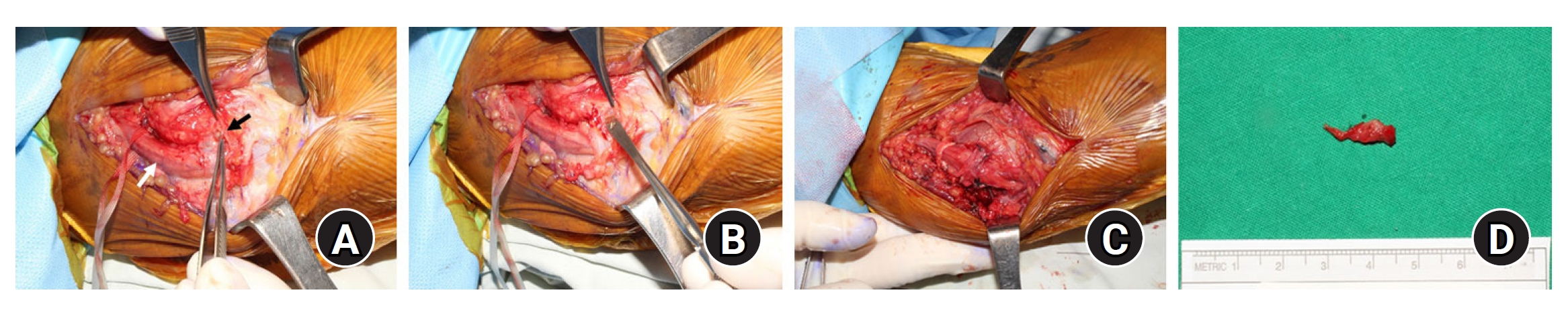
Intraoperative photography. (A) Swelling of ulnar nerve (white arrow) in cubital tunnel and Ganglion cyst (black arrow) compressing ulnar nerve in cubital tunnel. (B) Gel-like cyst compressing ulnar nerve. (C) Decompression and anterior transposition of ulnar nerve after removing the mass. (D) Excised ganglion cyst.
We confirmed that flexion and extension of the elbow did not cause subluxation of the ulnar nerve. The histopathological findings confirmed a ganglion mass consisting of a cystic wall and fibromyxoid tissue (Fig. 5). The patient began passive exercises after the surgery and showed improvement in hypoesthesia and weakness in the ring and little fingers 1 month postoperative. Moreover, ultrasound examinations performed 1 and 3 years after operation showed no recurrence.

Cyst wall composed of fibromyxoid tissue with fibrous wall (black arrow). H&E; (A) ×100, (B) ×400.
DISCUSSION
Ulnar nerve compression around the elbow due to an extrinsic lesion is usually caused by a tumor, hematoma, calcification, bone fragment, arthritic changes, or a cyst in the cubital tunnel. While ganglion cysts commonly develop in the wrist joint, ganglion cysts in the elbow associated with neurologic symptoms have also been reported [5]. However, CuTS caused by recurrent ganglion cysts is rare. We report a case of CuTS caused by ganglion cyst recurrence at the site of previous ganglion removal surgery.
In patients with CuTS, the prevalence of ganglion cysts has been reported to be 3% to 8%. During surgical excision of a ganglion cyst, removal of both the cyst and its stalk is necessary. If this is not done properly, postoperative recurrence is likely to occur. Komatsu et al. [6] reported a case series of nine patients with recurrent CuTS caused by ganglion cysts after surgical treatment of CuTS caused by a ganglion cyst. In our case, the patient had a history of ganglion cyst excision without neurologic symptoms. He complained of neurological symptoms in the upper extremity 4 years after the previous mass removal surgery. Before MRI, ganglion cyst was not suspected as a cause of CuTS. Since there is a strong association between osteoarthritis of the humeroulnar joint and medial elbow ganglia with CuTS, radiological examinations such as MRI and ultrasound should be used to determine whether it the condition is caused by space-occupying lesions [5].
There are numerous operative procedures for the treatment of CuTS including simple decompression, ulnar nerve transposition, and medial epicondylectomy, but most studies have shown no significant clinical differences between these methods [7]. Anterior transposition procedures can be grouped into subcutaneous anterior transposition, submuscular anterior transposition, and intramuscular anterior transposition. The disadvantages of submuscular anterior transposition are medial epicondylitis, longer recovery time, and possible nerve entrapment by a reattached muscle mass. Intramuscular anterior transposition risks surgical failure due to adhesion formation between the ulnar nerve and other fibrous anatomical structures [1,7]. A previous study reported that in cases involving a history of medial elbow surgery or distal humeral fracture, simple decompression with anterior transposition of the ulnar nerve was more effective than simple decompression due to significant amounts of scar tissue around the ulnar nerve [7]. Meanwhile, other studies also reported that anterior transposition of the ulnar nerve was more effective than simple decompression alone, with simple decompression alone leading to a high recurrence rate and increased intraneural pressure during elbow flexion after the procedure [7]. In our case, the MRI findings revealed a cyst at the site of the previous surgery. Entrapment of the ulnar nerve by mucous tissues was identified. The thickened ulnar nerve was dislocated during flexion because of an extensive incision on the Osborne ligament and the intermuscular septum of the FCU. Consequently, we performed decompression and additional subcutaneous anterior transposition of the ulnar nerve.
The postoperative recurrence rate of ganglion cysts varies between 0% and 31.2% [8], with an average recurrence period of 30 months postoperatively [9]. According to the literature, ganglion cysts may be suspected in cases involving a sudden exacerbation of CuTS symptoms within 2 months as well as medial elbow pain [5]. The patient in our case did not experience any discomfort after ganglion cyst excision. However, 4 years postoperative, he experienced a sudden onset of hypoesthesia in his hand and muscle weakness that persisted for 5 months. We identified a mass in the area of previous cyst excision. The histopathological findings confirmed that the mass had a higher proportion of fibrous tissue than typical ganglion cysts. Accordingly, we found that the mass was a recurrence of ganglion cyst with cicatricial changes resulting from previous surgery.
During excision of benign masses such as ganglion cysts, a sufficient explanation of the possibility of recurrence, secondary changes, and damage to surrounding structures due to scarring must be provided. Additionally, the joint capsule or aponeurosis with degenerative changes around the mass must be thoroughly removed together with the mass to prevent recurrence [9]. Our patient showed compressive neurological symptoms of CuTS caused by the recurrence of a ganglion cyst in the cubital tunnel in the area from which the original ganglion cyst was surgically excised. As is shown in our case, for patients with severe nerve dysfunction in which exact site of compression cannot be determined by electromyography alone, imaging studies are very useful for correcting preoperative diagnoses. When ulnar nerve palsy occurs, radiological examinations such as ultrasound and MRI must be performed in consideration of CuTS caused by a space-occupying lesion, in addition to compression caused by structural abnormalities. Moreover, the treatment should be determined in consideration of both simple decompression and anterior transposition.
Notes
Author contributions
Conceptualization: TK, WS. Data curation: TK. Formal Analysis: WS. Investigation: JH. Writing – original draft: TK, WS, JH. Writing – review & editing: TK, WS.
Conflict of interest
None.
Funding
None.
Data availability
None.
Acknowledgments
None.