Clin Shoulder Elb Search

CLOSE


Clin Shoulder Elb > Volume 27(1); 2024 > Article
Spek, Hoogervorst, Brink, Schoones, van Deurzen, and van den Bekerom: Ten technical aspects of baseplate fixation in reverse total shoulder arthroplasty for patients without glenoid bone loss: a systematic review

Abstract

The aim of this systematic review was to collect evidence on the following 10 technical aspects of glenoid baseplate fixation in reverse total shoulder arthroplasty (rTSA): screw insertion angles; screw orientation; screw quantity; screw length; screw type; baseplate tilt; baseplate position; baseplate version and rotation; baseplate design; and anatomical safe zones. Five literature libraries were searched for eligible clinical, cadaver, biomechanical, virtual planning, and finite element analysis studies. Studies including patients >16 years old in which at least one of the ten abovementioned technical aspects was assessed were suitable for analysis. We excluded studies of patients with: glenoid bone loss; bony increased offset-reversed shoulder arthroplasty; rTSA with bone grafts; and augmented baseplates. Quality assessment was performed for each included study. Sixty-two studies were included, of which 41 were experimental studies (13 cadaver, 10 virtual planning, 11 biomechanical, and 7 finite element studies) and 21 were clinical studies (12 retrospective cohorts and 9 case-control studies). Overall, the quality of included studies was moderate or high. The majority of studies agreed upon the use of a divergent screw fixation pattern, fixation with four screws (to reduce micromotions), and inferior positioning in neutral or anteversion. A general consensus was not reached on the other technical aspects. Most surgical aspects of baseplate fixation can be decided without affecting fixation strength. There is not a single strategy that provides the best outcome. Therefore, guidelines should cover multiple surgical options that can achieve adequate baseplate fixation.

INTRODUCTION

The worldwide incidence of reverse total shoulder arthroplasty (rTSA) has increased exponentially since the introduction of the first rTSA by Grammont et al. in 1987 [1-3]. Despite innovations in surgical technique and implant designs, rTSA-related complications occur in 19%–68% of patients [4-8]. The incidence of baseplate loosening following rTSA ranges from 1.2% to 11.7%, and it usually requires revision surgery [9-13]. Revision procedures following rTSA are associated with higher complication rates, worse functional outcomes, and decreased patient satisfaction compared to those of primary rTSA [13-16]. Therefore, it is important to prevent revision procedures in order to improve patient outcomes. However, achieving optimal glenoid baseplate fixation can be challenging. Several screw- and baseplate-related surgical fixation aspects, such as screw placement and baseplate characteristics, are believed to be critical for achieving optimal glenoid baseplate fixation.
Although various studies have assessed screw- and baseplate-related surgical fixation aspects in rTSA, there is still no consensus on how to achieve optimal glenoid-implant fixation in rTSA. Insight into optimizing glenoid-implant fixation in rTSA is important to reduce aseptic baseplate loosening requiring revision surgery, scapular notching, postoperative fractures, and suprascapular nerve (SSN) injury. Optimizing glenoid-implant fixation in rTSA may also improve patient outcomes. This review was performed with the goal of collecting the available evidence on the following ten technical aspects of baseplate fixation in rTSA: screw insertion angles; screw orientation; screw quantity; screw length; screw type; baseplate tilt; baseplate position; baseplate version and rotation; baseplate design; and anatomical safe zones.

METHODS

This systematic review process followed the Preferred Reporting Items for Systematic Reviews and Meta-Analyses (PRISMA) guidelines and was registered in the International Prospective Register of Systematic Reviews (identification number 245912) [17]. Ethical approval is not required for this type of study under Dutch law.

Search Strategy

The literature search was conducted in PubMed, Embase, Web of Science, Cochrane Central Library, and Emcare using a systematic search strategy (Supplementary Material 1) created by a librarian (JWS). The articles were selected from January 2000 to July 2022. The list of references was imported into EndNote (version X9) to remove duplicate articles. The references were subsequently exported to the web application Rayyan for study selection.

Study Selection

Three authors (RWAS, LAH, and RCB) independently screened the titles and abstracts before assessing the full texts for eligibility. Any discrepancies were resolved by discussion between the authors. Studies were included according to the following eligibility criteria: (1) inclusion of least three patients, all of whom were >16 years old; (2) analysis of at least one baseplate fixation aspect (screw insertion angle, screw orientation, screw quantity, screw length, screw type, baseplate tilt, baseplate position, baseplate version and rotation, anatomical safe zones) in rTSA; (3) data regarding clinical outcomes, biomechanical outcomes, and/or anatomical outcomes. We excluded studies of patients with: (1) glenoid bone loss (Walch type ≥B1); (2) bony increased offset-reversed shoulder arthroplasty; (3) rTSA with bone grafts; and (4) augmented baseplates. In addition, studies were excluded if the full text was unavailable, if data were not extractable or if it was any of the following study types: systematic review, meta-analysis, conference abstract, case report (defined as inclusion of less than three patients), expert opinion, or animal study. Finally, the reference lists of the retrieved articles were reviewed for additional articles (citation snowballing).

Critical Appraisal and Data Extraction

Methodological quality of the clinical and cadaver studies was appraised using the Critical Appraisal Skills Program (CASP) [18] and the Quality Appraisal for Cadaveric Studies (QUACS) checklists [19]. The CASP checklist was classified into low (<8 points) and high (≥8 points) levels of quality. The QUACS checklist was classified into poor (≤6 points), moderate (7–9 points), and good (≥10 points) levels of quality. Quality assessments, authors, year of publication, and data extraction of all included studies were independently extracted by three authors (RWAS, LAH, and RCB). Any discrepancies were resolved by discussion between the authors.

Statistical Analysis

Data was presented using descriptive statistics. Outcomes were not synthesized, as it was inappropriate to generate pooled effect sizes due to the between-study heterogeneity in methodology and outcomes. A brief summary of the reviewed material was presented after each section.

RESULTS

Literature Search

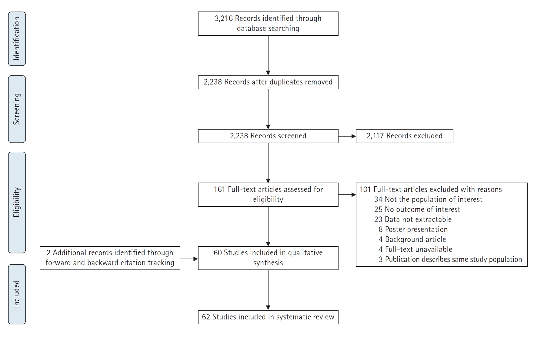
The literature search (Supplementary Material 1) identified 3,216 records. After removing duplicates, the titles and abstracts of 2,238 articles were screened. Thereafter, 161 full texts were assessed. Of these, 60 studies fulfilled the inclusion criteria. Another two studies were identified by reference checking; therefore, a total of 62 studies were included in the quality assessment (Fig. 1). The quality of clinical studies was low in 5 and high in 16 studies (Supplementary Material 2). The quality of cadaver studies was poor in zero, moderate in nine, and good in four studies (Supplementary Material 3). As zero studies were excluded after quality assessment, all 62 studies were suitable for analysis, including: 41 experimental studies (13 cadaver, 10 virtual planning, 11 biomechanical, and 7 finite element studies) and 21 clinical studies (12 retrospective cohort and 9 case-control studies).

Screw Insertion Angle

A finite element study in which four different diverging screw insertion angles (0°, 10°, 20°, and 30°) were tested showed that an increasing screw insertion angle resulted in reductions in the baseplate micromotions: 90–110 µm (screw insertion angle of 0°) and 48–59 µm (screw insertion angle of 30°) [20]. Meanwhile, a finite element study in which five different screw insertion angles (0°, 10°, 17°, 15°, and 34°) were analyzed showed that screw insertion angles of 17° provided the most optimal stress distribution on the humeral spacer [21]. A virtual planning study determined the optimal screw insertion angle according to two scenarios, as follows: (1) entire intraosseous screw trajectory, exiting in a “safe anatomical region” (i.e., avoiding injury to the SSN, which runs between the 2- and 8-o’clock positions with the right shoulder as reference); (2) in-out-in screw trajectory with penetration in the thickest cortical region regardless of anatomical structures [22]. The optimal screw insertion angles, according to this study, are summarized in Table 1. Additionally, because there are no important neurovascular structures located at the inferior scapular pillar, the authors emphasized that inferior screws should be angled into the inferior scapular pillar. The angles of the posterior and superior screws highly depend on surgeons’ preferences. For instance, superior screws could be directed laterally or inferiorly to the suprascapular notch, whereas posterior screws could be angled toward the lateral scapular spine area or to thin cortical areas (provided that the length is short). Compared with scenario 2, similar screw insertion angles for the inferior screws were found in a cadaver study (n=10), in which variable and fixed baseplates were used (Tables 1 and 2) [23]. To summarize, there were considerable differences in optimal screw insertion angles described in these experimental studies.

Screw Orientation

A cadaver study (n=20) reported no significant differences in baseplate micromotions measured by axial eccentric loading (0–300 N for 600 cycles) between the baseplates secured with divergent or parallel oriented screws (2.0 µm, standard error: 0.7 vs. 4.0 µm, standard error: 1.5, respectively) [24]. Additionally, a biomechanical study using cycling loading (500 N for 1,000 cycles) observed no significant differences in baseplate micromotions among baseplates secured with neutral or divergent oriented screws, as follows: inferior 247±22 and 193±23 µm; superior 121±17 and 108±18 µm; anterior 180±16 and 153±17 µm; posterior 188±22 and 148±23 µm [25]. Contrarily, a finite element study showed that baseplates secured with divergent oriented screws demonstrated less baseplate micromotions than did those secured with convergent oriented screws [26]. Likewise, another finite element study using compressive and shear load of 750 N demonstrated that divergent oriented screw fixation resulted in less baseplate stress and displacement than did baseplates secured with convergent or parallel oriented screws (Table 3) [27].
In summary, two out of four experimental studies suggested that baseplates should be secured with divergent oriented screws. In contrast, two experimental studies found no differences in baseplates micromotions while using different screw orientations.

Screw Quantity

A cadaver study (n=4) reported reduced baseplate micromotions among baseplates secured with four screws when compared to those secured with two screws (18.3±5.9 vs. 35.0±14.9 µm; P=0.01, respectively) [28]. Additionally, a biomechanical study in which baseplates were constructed with two, four, or six screws reported more displacement both pre- and post-cyclic loading (750 N for 10,000 cycles) in baseplates secured with two screws than in baseplates secured with four or six screws (two screws 116±36 and 125±44 µm; four screws 82±22 and 91±23 µm; six screws 92±20 and 108±42 µm, pre- and post-cyclic loading, respectively). However, no differences were observed between four versus six screws (P=0.18 and P=0.18, pre- and post-cyclic loading, respectively) [29]. Furthermore, a cadaver study (n=10) analyzed the added value of the posterior screw by measuring the amount of vertical displacement of the glenoid component during cyclic loading (750 N for 50,000 cycles); this group found a three-fold higher rate of glenoid loosening in baseplates without posterior screws compared to those with posterior screws [30]. Contrarily, no significant differences in baseplate displacement were identified in another cadaver study (n=12) using cycling loading (650–1,000 N for 100 cycles in superior directions followed by 100 cycles in anterior-posterior directions). This group was primarily looking for mean differences in displacements between two and four screws constructs of: anterior 42 µm; posterior 41 µm; superior 13 µm; and inferior 14 µm [31]. Similar outcomes were reported in a biomechanical study using cyclic loading of 500 N for 1,000 cycles. This group found mean displacements between two and four screws, respectively, as follows: inferior 235±23 and 205±22 µm; superior 130±18 and 99±17 µm; anterior 180±16 and 155±16 µm; posterior 187±23 and 149±22 µm [25].
A case-control study (n=3,180) including a biomechanical model using compressive loading (10 mm/min) reported that superior screw insertion within four-screw constructs was associated with: a higher incidence of scapula body fractures (4.4% vs. 0.0%, superior screws yes/no, respectively); and a lower load to failure (1,077 N vs. 1,970 N, superior screws yes/no, respectively) [32]. Similar findings were reported in a case-control study (n=4,125) with a minimum follow-up of 2 years. In this study, patients with acromial and/or scapular spine fractures had more baseplate screws than did those without fractures (4.05±0.51 vs. 3.83±0.79, respectively; P=0.02) [33]. In contrast, another retrospective study (n=105) showed that utilizing three or four screws (vs. only two screws) did not increase the odds of minor or major radiographic changes (Table 4) [34].
To summarize, three out of five experimental studies demonstrated that baseplates should be secured with four screws. However, two out of three clinical studies reported higher occurrences of scapular and/or acromial fractures as the number of baseplate screws increases.

Screw Length

Five experimental studies analyzed the optimal screw lengths based on anatomical structures and/or maximum cortical fixation (Table 5) [22,23,35,36]. A finite element analysis study reported a 30% reduction rate in the baseplate micromotion using 30-mm instead of 16-mm screws [20]. Likewise, a biomechanical study reported lower baseplate micromotions after cycling loading (500 N for 1,000 cycles) among baseplates secured with 36-mm compared to 18-mm screws. This group described mean displacements with 36-mm vs. 18-mm screws, respectively, as follows: inferior 258±23 and 182±22 µm; superior 114±18 and 115±17 µm; anterior 190±17 and 143±16 µm; posterior 182±23 and 154±22 µm [25]. Additionally, a biomechanical study observed that the lowest baseplate displacements (both pre- and post-cyclic loading 750 N for 10,000 cycles) occurred in baseplates secured with 46-mm screws, followed by baseplates secured with 30- and 18-mm screws (46-mm screws: 74±15 and 73±8 µm; 30-mm screws: 101±12 and 111±16 µm; 18-mm screws: 115±39 and 140±45 µm, pre- and post-cyclic loading, respectively) [29]. The last cadaver study (n=7) described the use of long screws and showed that outside-in screws, as well as long screws, are risk factors for scapular fractures [37].
A case-control study reported no significant differences in the screw length of posterior and superior screws between patients with (n=53) or without (n=212) scapular spine fractures (23 vs. 22 mm, respectively) [38]. Similarly, a case-control study assessed the relationship between increasing screw length and the occurrence of acromial fractures, but did not find an association [39]. Additionally, a retrospective cohort study (n=82) assessed the incidence of glenoid penetration and found that all posterior screws with a length >20 mm (n=82) penetrated the glenoid vault (Table 6) [40].
Taken together, seven experimental studies reported benefits of fixating the baseplates with screws that are at least 30 mm in length. Two clinical studies reported no significant differences in screw length between patients with or without scapular fractures. One clinical study strongly advised against using >20 mm posterior screws.

Screw Type

The authors of a biomechanical study suggested using at least two locking screws, because they require a higher load to failure (2,153±115 N) compared to constructs with four non-locking screws (1,832±35 N) (P<0.01) [41]. Another biomechanical study demonstrated that baseplates secured with four locking screws had less baseplate micromotion than did those secured with four non-locking screws (P=0.02) [42]. Contrarily, another biomechanical study tested four screw combinations (1 locking screw vs. 3 non-locking screws, 2 vs. 2, 3 vs. 1, and 4 vs. 0) using cyclic loading (750 N for 10,000 cycles). This group reported no significant differences in baseplate micromotions after cycling loading between the following combinations (reported with their mean micromotions): 1 locking screw (97.1±47.2 µm); 2 locking screws (76.7±34.5 µm); 3 locking screws (72.4±15.3 µm); 4 locking screws (68.1±15.3 µm) [43]. Another biomechanical study analyzed the use of locking versus non-locking screws from another perspective. These authors concluded that if the central element punctured well into the cortical bone, non-locking anterior and posterior screws were sufficient. On the contrary, if the central element was too short, the anterior-posterior screws were required to have a locking function [44]. Furthermore, a cadaver study (n=10) compared the position of locking screws (superior-inferior locking screws with anterior-posterior compression screws versus anterior-posterior locking screws with superior-inferior compression screws) and found no difference in micromotion between these different positions (Table 7) [25].
In summary, two out of four biomechanical studies recommended securing baseplates with at least two locking screws. One biomechanical study showed that locking screws were particularly important if the central peg did not puncture into the cortical bone. The cadaver study demonstrated that the position of the locking screws does not improve the fixation strength.

Baseplate Tilt

Previous experimental studies that described the relationship between tilt and baseplate stress and impingement are contradicting [27,45-48]. A retrospective cohort (n=146) with a mean follow-up of 21 months reported that scapular notching significantly decreased when an inferior tilt was used. However, baseplate tilt angles did not affect the range of motion (ROM) or functional-, pain-, and satisfaction-scores [49]. Furthermore, another retrospective cohort (n=71) found no differences in the grade or incidence of scapular notching at a minimum of 12 months of follow-up between baseplates secured in neutral (0°) and inferior (–10° or –15°) tilt: 76.7% vs. 60.7%, respectively (P=0.08) [50]. Additionally, a case-control study (n=136) concluded that baseplate inclination was not related to the likelihood of developing implant instability [51]. A retrospective cohort (n=105) reported that superior tilt was associated with increased risks of scapular notching and signs of loosening (odds ratio [OR]: 2.52 and OR: 8.92, respectively) [34]. However, another retrospective study (n=154) described no significant difference in postoperative ROM, patient-reported outcomes (PROMS), scapular notching, and heterotopic ossification between inferior, neutral, and superior (up to 6°) glenoid baseplate inclination [52]. Comparable results were described in a retrospective case-control study (cases=34 and controls=102); the final prosthetic glenoid inclination, as well as the change in glenoid inclination, had no influence on the risk of prosthetic instability [51]. Additionally, a cohort study (n=61) concluded that glenoid inclination had no significant influence on clinical outcomes at a minimum follow-up of 2 years [53]. Contrarily, another case-control study (n=33) reported an association between baseplate tilt and implant stability, as follows: –10.2° tilt in stable versus 8.3° in unstable implants (P=0.01) [54]. Likewise, another case-control study (n=97) reported a 13% instability rate at a mean follow-up of 47 months, and the only factor found to be associated with it was superior tilt: OR: 1.15, P=0.01) (Table 8) [55].
To summarize, the evidence is inconclusive to formulate guidelines with regard to baseplate tilt. Nevertheless, all prior studies have recommended against superior tilt.

Baseplate Position

Despite contradictory results, most experimental studies preferred an inferior position of the baseplate to increase the peak load failure and improve rotation [56-63]. A retrospective cohort study (n=54) showed that patients with scapular notching had higher positioned baseplates (as measured from the baseplate’s inferior aspect to the inferior rim of the glenoid) than did those without scapular notching (2.8±3.3 vs. 0.6±2.0 mm, P=0.03, respectively) [64]. Another retrospective cohort study (n=77) demonstrated that patients with inferior notching had higher peg-glenoid rim distances than did those without inferior notching (24.7±3.0 vs. 20.1±2.5 mm, P<0.001, respectively) [65]. Furthermore, a retrospective cohort (n=151) concluded that patients with scapular notching and/or osteophyte formation had higher positioned baseplates than did those without either scapular notching or osteophyte formation (20.3 vs. 19.1 mm, respectively) [66]. Moreover, a cohort study reported significantly longer peg glenoid rim distances and shorter sphere bone overhang distances in 13 patients with scapular notching as compared to 58 patients without scapular notching (24.8±1.6 and 2.6±0.5 mm vs. 21.9±1.9 and 5.8±1.9 mm, respectively). However, no significant differences were found in shoulder function and active ROM between the two patient groups at the last follow-up (37.0±3 months) [67]. A retrospective review (n=105) with a minimum follow-up time of 5 years found an increased risk of severe scapular notching that was mainly associated with a high (the glenosphere grazed the inferior edge of the glenoid, OR: 2.68) or excessively high (the glenosphere was beyond the inferior edge of the glenoid, OR: 7.55) position [34]. A retrospective cohort (n=97) analyzing glenoid components with ≥3.5 mm of inferior overhang versus flush glenoid components described a significantly lower rate of radiographic notching (37% vs. 82.5%, respectively), better clinical outcomes, and higher subjective shoulder value if the glenoid component had at least 3.5mm of inferior overhang as compared to a flush glenoid component [68]. Contrarily, another retrospective cohort (n=147) concluded that inferior positioned baseplates were associated with increased rates of scapular notching. However, baseplate positions were not associated with the incidence of revision surgery (28.9 vs. 25.2 mm, P=0.17, revision yes/no, respectively) (Table 8) [69]. In summary, the majority of the experimental and clinical studies reported benefits of fixating baseplates inferiorly.

Baseplate Version and Rotation

A biomechanical study analyzed the differences between five different glenosphere positions (20° retroversion, 10° retroversion, neutral position, 10° anteversion, and 20° anteversion) on implant stability and concluded that baseplates should be secured in anteversion or a neutral position to attain the highest stability ratio [70]. A finite element analysis showed that a neutral glenoid component produced the greatest impingement-free ROM, as compared to 5° anteversion and 5°, 10°, and 20° retroversion [71].
A case-control study (including patients with scapular spine fractures (n=53) and controls without scapular spine fractures (n=212), reported no significant differences in baseplate anteversion between the two groups [38]. The baseplate was anteverted in 20% of the cases and in 17.6% of the controls [38]. According to a finite element analysis model, the baseplate retroversion does not need to be corrected to <10° to provide good initial fixation. Instead, it can withstand the initial stresses and micromotion up to 25° of retroversion (Table 8) [72]. A virtual planning study was the only study to examine the influence of internal baseplate rotation; the group reported that 11° of internal rotation from the 12 o’clock position resulted in the strongest superior screw fixation [73].

Baseplate Design

Curved back or flat back?

A biomechanical study reported no differences in shear displacement both pre- and post-cycling loading (750 N for 10,000 cycles) between curved-back and flat-back baseplates [74]. However, a virtual planning study showed better bone contact surface area in curved-back baseplates when compared to flat-back baseplates (P=0.01) despite the fact that flat-back implants had better screw puncture and less bone removal during fixation than did curved-back baseplates (P=0.03 and P=0.01, respectively) [75]. Another virtual planning study analyzed the amount of bone removed during reaming in three different baseplate designs (two curved-back and one flat-back baseplates). This group reported that the amount of bone removal was the highest among 26-mm curved-back baseplates, followed by 29-mm flat-back and 25-mm curved-back baseplates (Table 9) [60].

Circular or oval?

Only one study examined the outcomes of circular versus oval baseplates. This biomechanical study showed that circular baseplates had more shear displacement in both the superior-inferior and anterior-posterior directions both pre- and post-cyclic loading (750 N for 10,000 cycles) than did oval baseplates (Table 9) [73].

The smaller the better?

A cadaver study (n=5) demonstrated that 25-mm baseplates, when compared to 29-mm baseplates, resulted in less baseplate micromotion at the inferior third of the glenoid-glenosphere interface, a smaller shoulder adduction deficit, and a greater impingement-free ROM [76]. However, no differences in baseplate displacement between 25- and 24-mm baseplates were found in a biomechanical study using cyclic loading (750 N for 10,000 cycles) [29].
One retrospective cohort (n=11) analyzed the outcomes of a 25-mm baseplate in a cohort of relatively short patients (mean length: female, 156±8 cm; male, 171±2 cm). Despite a high rate of scapular notching (82%), outcomes at 3 years of follow-up were successful, including: no revision procedures, no radiographic evidence of implant loosening and acceptable ROM, PROMs, and strength (Table 9) [78].
In summary, one cadaver study reported superior outcomes of 25-mm and 29-mm baseplates, whereas one biomechanical study found no differences in baseplate displacement between 25- and 24-mm baseplates. One retrospective cohort demonstrated acceptable outcomes of 25-mm baseplates.

The 2- or 1-peg design?

A case-control study (n=85) in which 2- and 1-peg baseplates were compared reported a lower rate of scapular notching, polyethylene induces osteolysis, and metal screw contact when using 2-peg baseplates [77]. However, the amount of baseplate micromotion following cyclic loading (750 N from 0–100,000 cycles) did not differ between the two constructs (47 and 43 µm, 2- and 1-peg baseplates, respectively).

Central peg of central screw fixation?

A biomechanical study concluded that central screw fixation resulted in less baseplate micromotion than did central peg fixation. Also, the central elements that puncture the cortical bone result in less micromotion than do the shorter ones, which do not reach the cortex [44].

Anatomical Safe Zones

A virtual planning study (n=56) described a danger zone to assist surgeons to avoid SSN injury and revealed that the danger zone of the superior screw was located between the 2- and 8-o’clock positions (using the 12 o’clock position of the right glenoid as reference) [79]. The posterior screw touched the neurovascular structures in 33% of specimens in a cadaver study (n=10) [36]. Additionally, another cadaver study (n=10) showed that the superior and posterior screws posed the most risk to the SSN, with a 40% chance of touching the SSN [80]. Even higher rates of SSN engagement were reported in another cadaver study (n=12) in which the superior screw touched the SSN in 8 (66%) and the posterior screw in 6 specimens (50%). This group also concluded that overly long screws pose a serious risk for SSN injury and advised <2 mm penetration for superior or posterior screws [81].
One retrospective study (n=82) concluded that 13% of superior screws and 65% of posterior screws penetrated the glenoid vault. Among the superior screws, 64% had a high-risk of iatrogenic SSN neuropathy (screw tip placed within 5mm of the nerve), while only 6% of posteriorly inserted screws carried the same risk. Comparison analysis showed no difference in PROMs between the high- and low-risk (screw tip placed >5 mm of the nerve) penetrations (Table 10) [40].
In summary, four experimental studies proved that far-cortex penetration by the superior and posterior screw should be avoided to minimize the likelihood of neurovascular injuries. One experimental study described a danger zone of the superior screw between the 2- and 8-o’clock positions (using the 12 o’clock position of the right glenoid as reference). However, a clinical study showed that screw penetrations close to the SSN (high-risk) did not portend poorer clinical outcomes compared to screw penetrations far away from the nerve (low-risk).

DISCUSSION

As universal guidelines on baseplate fixation are lacking, this review sought to provide a narrative overview of the currently available evidence on ten baseplate fixation aspects in rTSA. So far, it can be deducted that: (1) Optimal screw insertion angles are unknown. Therefore, until more evidence is gathered, surgeons should focus on adequate screw puncture in anatomical safe zones and driving the inferior screw into the inferior scapular pillar; (2) Finite element studies advise the use of divergent screw patterns only, while cadaver studies conclude that both parallel or divergent patterns are sufficient for adequate stability; (3) An increasing number of screws leads to a reduced baseplate micromotion, but it is also associated with a higher risk of acromial fractures; (4) Posterior screws should be shorter or equal to 20 mm, while other screws should be 30 mm or longer; (5) If the central element does not puncture cortical bone, peripheral anterior and posterior locking screws are recommended. It is noteworthy that apart from one study, there seems to be a benefit of using at least some locking screws in baseplate constructs; (6) The optimal baseplate tilt is unknown, but the baseplate is best secured inferiorly in either slight anteversion or a neutral position; (7) There is no consensus on the best type of baseplate; and (8) Far cortex penetration should be avoided. Due to the lack of (large) clinical studies, methodological- and outcome-heterogeneity, these conclusions should be considered preliminary clinical advice.
Although this review is a collection of the best evidence available, several limitations should be acknowledged. First, the majority of the included studies were experimental studies. Therefore, their shortcomings, when compared to clinical studies, should be taken into consideration [82-84]. Furthermore, the biomechanical and virtual planning studies did not consider additional factors that are likely to affect rTSA biomechanics (e.g., stabilizing effects of ligaments, rotator cuff muscles, patients’ daily activities, and anatomical variations). Second, although no cadaver studies were judged as “poor” on quality assessment, only four out of 14 cadaver studies were assessed as having “good” quality. Third, most studies underreported their statistical results such as confidence intervals and/or standard deviations. Fourth, most included studies had methodological inconsistencies such as a lack of power, small sample size, short follow-up duration, and heterogenic outcomes. Furthermore, due to the between-study heterogeneity of the outcomes and patient characteristics, it was inappropriate to synthesize the outcomes to generate pooled effect sizes. In addition, this review only focused on studies including patients without glenoid bone loss, because baseplate fixation in patients with glenoid bone loss requires different fixation techniques compared to patients without glenoid bone loss [85]. Several clinical studies had to be excluded because they analyzed patients with and without glenoid bone loss, and data from these distinct patient groups were not extractable. A last limitation is that some studies used baseplate micromotion as the primary outcome and concluded on superiority relative to the comparison group, despite the fact that the amount of micromotion was far below the commonly accepted threshold of osseointegration failure (150 µm). Still, how much micromotion will result in clinical adverse events such as baseplate loosening and revision surgery remains unclear [86].

CONCLUSIONS

Most surgical aspects of baseplate fixation can be decided without affecting fixation strength. There is not a single strategy that provides the best outcome. Therefore, guidelines should cover multiple surgical options that can achieve adequate baseplate fixation. This also implies that surgeons can opt for their desired fixation method during surgery.

NOTES

Author contributions

Conceptualization: RWAS, LAH, DFPD, MPJB. Formal analysis: RWAS, LAH, RCB. Methodology: RWAS, LAH, JWS, DFPD, MPJB. Resources: MPJB. Supervision: RCB, JWS, DFPD, MPJB. Validation: MPJB. Visualization: RWAS, LAH. Writing–original draft: RWAS, LAH. Writing–review & editing: RWAS, LAH, RCB, DFPD, MPJB.

Conflict of interest

The first author (RWAS) received funding as indicated in "Funding" section. No other potential conflicts of interest relevant to this article were reported.

Funding

The first author (RWAS) has received payments during the study period including <$10,000 from Anna Fonds NOREF (Mijdrecht, the Netherlands), <$10,000 from Michaël-van Vloten Fonds (Rotterdam, the Netherlands), and an amount between $10,000-100,000 from Prins Bernhard Cultuurfonds (Amsterdam, the Netherlands) and Flinders Foundation (Adelaide, Australia).

Data availability

None.

Acknowledgments

None.

SUPPLEMENTARY MATERIALS

Supplementary materials can be found via https://doi.org/10.5397/cise.2023.00493.
Supplementary Material 1.
cise-2023-00493-Supplementary-Material-1.pdf
Supplementary Material 2.
cise-2023-00493-Supplementary-Material-2.pdf
Supplementary Material 3.
cise-2023-00493-Supplementary-Material-3.pdf

Fig. 1.
Flowchart of included studies.
cise-2023-00493f1.jpg
Table 1.
Optimal screw insertion angles in degrees
First author (year) Definition Superior screw Posterior screw Anterior screw Inferior screw
DiStefano (2011) [22] Intraosseous through cortical bone and exits a ‘‘safe region’’ (based on anatomical structures) Penetrates the thickest cortical region regardless of anatomical structures 9±3 (S/I) –29±8 (S/I) –16±5 (S/I) –16±7 (S/I)
–2±5 (A/P) 3±7 (A/P) –14±4 (A/P) 5±4 (A/P)
28±6 (S/I) 23±4 (S/I) –16±5 (S/I) –19±6 (S/I)
10±6 (A/P) –3±6 (A/P) –14±4 (A/P) 4±4 (A/P)
Humphrey (2008) [23] Maximized screw length, accomplished far cortical fixation, and attained screw purchase in good bone stock 19 (S), 5 (I) 14 (I), 7 (A)
Variable-angle baseplate; fixed-angle baseplate 20(S), 20 (I) 20 (I), 20 (A)

Values are presented as mean±standard deviation.

S: superior, I: inferior, A: anterior, P: posterior.

Table 2.
Overview of included studies for screw insertion angle
First author (year) Design Brand Base plate Peg Screw Loading Conclusion
Humphrey (2008) [23] Cadaver (n=10) Aequalis (Tornier) - - 4 Screws - Table 1
Basat (2018) [21] Finite element Delta Xtend (Depuy) 36-mm diameter 6-mm diameter, 13-mm length 4 Screws, - Screw insertion angle s of 17° provided the optimal stress distribution on the humeral spacer.
4.5-mm diameter,
24-mm length
Hopkins (2008) [20] Finite element Delta III (Depuy), - - 4 Screws, 756 N axial superior Increasing the screw insertion angle resulted in less BP motion.
RSP neutral (Encore Medical), RSP reduced (Encore Medical) 3.5- or 5-mm diameter, 16- or 30-mm length
DiStefano (2011) [22] Virtual planning Aequalis (Tornier) 29-mm diameter 8-mm diameter 4 Screws - Table 1

-: not reported, BP: baseplate.

Table 3.
Overview of included studies for screw orientation
First author (year) Design Brand Base plate Peg Screw Loading Conclusion
Lung (2019) [25] Biomechanical Delta Xtend (DePuy) - 13.5- or 23.5-mm length 2 or 4 Screws, 18- or 36-mm length 500 N for 1,000 cycles Divergent versus neutral screw orientation: no differences in base plate motion
Abdic (2021) [24] Cadaver (n=20) Aequalis (Tornier) 29-mm diameter 8-mm diameter 4 Screws, 4.5-mm diameter 0–300 N for 600 cycles Divergent versus parallel screw orientation: no differences in base plate motion
Denard (2017) [27] Finite element Univers Revers (Arthrex) 24-mm diameter 6.5-mm diameter, 15-mm length 2 Screws, Compressive and shear load 750 N Divergent screws resulted in less base plate stress and displacement compared to parallel and convergent orientations.
4.4-mm diameter,
24-mm length
Yang (2013) [26] Finite element Aequalis (Tornier) - - - - Divergent screw orientation resulted in less base plate motion compared to convergent orientations.

-: not reported.

Table 4.
Overview of included studies for screw quantity
First author (year) Design Brand Base plate Peg Screw Loading Conclusion
Lung (2019) [25] Biomechanical Delta Xtend (DePuy) - 13.5- or 23.5-mm length 2 or 4 Screws; 18- or 36-mm length 500 N for 1,000 cycles No differences in base plate motion between 2- and 4-screw constructs
Roche (2019) [29] Biomechanical Equinoxe (Exactech) 25- or 24-mm diameter - 2, 4, or 6 Screws, 750 N for 10,000 cycles Using 4 or 6 instead of 2 screws resulted in less base plate motion. No differences between 4 versus 6 screws
4.5-mm diameter,
18- or 30- or 46-mm length
Elwell (2017) [28] Cadaver (n=4) - 25-mm diameter 8-mm diameter, 2 or 4 Screws, Direct force 686 N Using 4 instead of 2 screws resulted in less base plate motion.
15-mm length 4.5-mm diameter
Hoenig (2010) [30] Cadaver (n=10) Aequalis (Tornier) - - 3 or 4 Screws, 750 N for 50,000 cycles Absence of posterior screws (3 screws) resulted in higher rates of glenoid loosening than base plates with posterior screws (4 screws).
22- or 29-mm length
James (2013) [31] Cadaver (n=12) Aequalis (Tornier) - - 2 or 4 Screws 650–1,000 N for 100 cycles superior followed by 100 cycles anterior-posterior No differences in base plate motion between 2- and 4-screw constructs
Routman (2020) [33] Case-control (n=4,125) Equinoxe (Exactech) - - - - Increased risk of scapular and/or acromial fractures when using more base plate screws
Kennon (2017) [32] Case-control (n=318) including a biomechanical model Equinoxe (Exactech) - - 3 or 4 Screws, Compressive load 10 mm/min Presence of superior screws (4 screws) resulted in higher scapular fracture rates and lower load to failure than base plates without superior screws (3 screws).
4.5-mm diameter,
18- or 38-mm length
Lopiz (2021) [34] Retrospective cohort study (n=105) Delta III (DePuy), Delta Xtend (DePuy), Lima SMR (LimaCorporate) - Standard - - 3 or 4 versus 2 screws did not yield higher odds for minor or major radiographic changes.
Table 5.
Optimal screw lengths in millimeters
First author (year) Definition Superior Posterior Anterior Inferior
Codsi (2007) [35] Maximum screw length 29 75
DiStefano (2011) [22] Intraosseous through cortical bone and exits a ‘‘safe region’’ (based on anatomical structures)> 35 and 36 19 and 37 29 and 29 34 and 35
Penetrates the thickest cortical region regardless of anatomical structures
Hart (2013) [36] Maximized screw length, without damaging neurovascular structures 30 15 13 28
Humphrey (2008) [23] Maximized screw length, accomplished far cortical fixation, and attained screw purchase in good bone stock 36 and 33 47 and 43
Variable-angle baseplate, fixed-angle baseplate
Table 6.
Overview of included studies for screw length
First author (year) Design Brand Base plate Peg Screw Loading Conclusion
Lung (2019) [25] Biomechanical Delta Xtend (DePuy) - 13.5- or 23.5-mm length 2 or 4 Screws, 500 N for 1,000 cycles Less base plate motion when using 36-mm screws when compared to 18-mm screws
18- or 36-mm length
Roche (2019) [29] Biomechanical Equinoxe (Exactech) 24- or 25-mm diameter - 2, 4, or 6 screws, 750 N for 10,000 cycles Lowest base plate motion when using 46-mm screws, followed by 30-, and 18-mm screws
4.5-mm diameter, 18- or 30- or 46-mm length
Hart (2013) [36] Cadaver (n=10) RSP Encore (DJO Surgical) - 6.5-mm diameter 4 Screws, - Table 5
5-mm diameter
Humphrey (2008) [23] Cadaver (n=10) Aequalis (Tornier) - - 4 Screws - Table 5
Hopkins (2008) [20] Finite element Delta III (DePuy); RSP neutral (Encore Medical); RSP reduced (Encore Medical) - - 4 Screws, 756 N axial superior loading Using 30-mm screws instead of 16-mm screws resulted in 30% less base plate motion.
3.5- or 5-mm diameter,
16- or 30-mm length
Codsi (2007) [35] Virtual planning - - - 4-mm diameter - Table 5
DiStefano (2011) [22] Virtual planning Aequalis (Tornier) 29-mm diameter 8-mm diameter 4 Screws - Table 5
Otto (2013) [38] Case-control (n=265) - - - 14–30-mm length - No differences in screw length between patients with or without scapular fractures
Cho (2021) [39] Case-control (n=787) Aequalis (Tornier), Equinoxe (Exactech), TM Reverse (Zimmer), Ascend Flex (Tornier), Comprehensive Reverse (Biomet), RSP (DJO Surgical), SMR (Lima), Delta Xtend (DePuy), Anatomical shoulder (Zimmer) Several Several Several - Screw size is not correlated with acromion fractures.
Jang (2022) [40] Retrospective cohort (n=82) RSP (DJO Surgical), comprehensive (Biomet), Aequalis ascend flex (Wright Medical) 30-, 20-, 25-mm diameter, respectively - Length superior screw: 28±4 mm (15–35), posterior screw: 18±3 mm (14–30) - All posterior screw lengths >20 mm penetrated the glenoid vault
Table 7.
Overview of included studies for screw type (locking vs. non-locking)
First author (year) Design Brand Base plate Peg Screw Methods Conclusion
Chebli (2008) [41] Biomechanical Delta (DePuy) - - 4 Screws, 200 N preloaded, followed by 30 mm/sec Higher load to failure when using locking screws instead of non-locking screws
36-mm length
Formaini (2017) [43] Biomechanical RSP (DJO Global) - 6.5-mm diameter 4 Screws, 750 N for 10,000 cycles No differences in base plate motion when using 1 locking screw vs. 3 non-locking screws; 2 vs. 2; 3 vs. 1; 4 vs. 0
3.5- or 5-mm diameter,
22-mm length
Harman (2005) [42] Biomechanical Delta III (DePuy); RSP (Encore Medical) 23- or 27-mm diameter 16-mm length 4 Screws, 756 N for 1,000 cycles Less base plate motion when using locking screws
3.5- or 5-mm diameter
Torkan (2022) [44] Biomechanical Delta XTEND (DePuy) - 13.5-mm central peg vs. 23.5 mm central screw (diameter: 6.5 mm) 2 Peripheral screws (anterior/ posterior), non-locking vs. locking Compressive loading 500 N, 1 Hz, 1,000 cycles Anterior and posterior holes can be non-locking screws if the central peg purchases the cortical bone.
Abdic (2021) [24] Cadaver (n=10) Aequalis (Tornier) 29 mm 8-mm-diameter central post 2 Compression and 2 locking screws (AP vs. SI) Compressive loading and cyclic test AP vs. SI locking screw position: similar fixation strength

AP: anteroposterior, SI: superoinferior.

Table 8.
Overview of included studies for baseplate tilt, position, version, and rotation
First author (year) Design Brand Base plate Peg Screw Loading Conclusion
Favre (2010) [70] Biomechanical Delta III (DePuy) - - - Constant load of 40 N, constant displacement of 10 mm/sec Anteversion or neutral position resulted in highest implant stability ratios.
Gutiérrez (2007) [46] Biomechanical RSP (Encore Medical) 25-mm diameter - 4 Screws - Inferior (–15°) tilt resulted in less base plate motion than neutral (0°) or superior (15°) tilt.
Fukuta (2021) [56] Biomechanical Trabecular Metal Reverse Shoulder (Zimmer) 28 mm 15×8 mm 2 Locking - Shifting the center of the baseplate slightly anterior to the anatomic center avoids perforation of the scapular neck in small glenoids.
Huish (2021) [57] Biomechanical Aequalis (Tornier) 29 mm - - 3D modelling Increased glenosphere overhang improved internal rotation. Glenoid lateralization alone did not.
Chae (2016) [45] Cadaver (n=5) Aequalis (Tornier) 29-mm diameter - 4 Screws Cyclic loading 480–686 N Inferior (–10°) tilt resulted in more base plate motion and higher bone stress than neutral (0°) tilt.
Pauzenberger (2019) [59] Cadaver (n=36) Univers Revers, 24.4–30.4-mm diameter 6.5-mm diameter; 3 Screws, - Inferior position resulted in higher peak load at failure than superior position.
II (Arthrex) 15-, 20-, or 4.5- or 6.5-mm diameter,
25-mm length 24-48-mm length
Wright (2015) [62] Cadaver (n=24) Comprehensive (Biomet) - 6.5-mm diameter 4 Screws, 30 N for 5 minutes, followed by 10 mm/min Inferior position resulted in decreased elongation and displacements of the deltoid.
6.5-mm diameter
Denard (2017) [27] Finite element Univers Revers (Arthrex) 24-mm diameter 6.5-mm diameter; 15-mm length 2 Screws, Compressive and shear load 750 N Inferior (–10°) tilt resulted in less base plate stress and displacement than neutral (0°) or superior (10°) tilt.
4.5-mm diameter,
24-mm length
Zhang (2019) [63] Finite element Grammont rTSA (not specified) - - 4 Screws - Inferior position resulted in increased base plate motion.
Friedman (2021) [72] Finite element Equinox (Exactech) - - Four 4.5-mm compression screws Different runs, including maximum stress To provide good initial fixation, retroversion does not need to be corrected to <10° and can withstand the stress and micromotion up to 25° of retroversion.
Permeswaran (2017) [71] Finite element Aequalis (Tornier) - - - Constand load of 40 N during different degrees of extension/flexion and ER/IR Neutral glenoid component version was associated with the lowest incidence of subluxation.
Ingrassia (2019) [47] Virtual planning Aequalis (Tornier) - - - - Inferior (–15°) tilt resulted in higher ROM and implant stability than neutral (0°) tilt
Roche* (2013) [60] Virtual planning Delta III (DePuy), 25- or 26- or 29-mm diameter 8-mm diameter; 4 Screws, - Inferior tilt resulted in more bone removal.
RSP (DJO), 16.5-mm length 4.5-mm diameter,
Equinoxe (Exactech) 30-mm length
Stephens (2015) [73] Virtual planning Comprehensive mini base plate (Biomet) 25-mm diameter 6.5-mm diameter 4 Screws, - 11° Internal rotation (using the 12 o’clock position of the right glenoid as reference) resulted in maximal base plate fixation strength.
<15–35-mm length
Jeong (2021) [58] Virtual planning Comprehensive (Zimmer-Biomet), Coralis (Corentec), DELTA XTEND (DePuy-Synthes), Trabecular Metal (Zimmer-Biomet) 30-mm diameter 6.5-mm diameter, 35-mm length - Central peg was inserted on 49 points and overlapped volume was determined. Optimal insertion site of the baseplate is located 2 mm inferiorly and posteriorly to the point where the vertical and horizontal axes of the glenoid intersect.
Patel (2021) [48] Virtual planning Perform Reverse 25-mm diameter - - - Inferior tilted base plates lead to a decrease in impingement free external rotation and adduction compared to neutrally positioned baseplates.
Tashiro (2022) [61] Virtual planning Equinoxe (Exactech) - 7.5-mm diameter, 16.6-mm length - - Lower risk of peg penetration when baseplate is positioned anterior to the long axis of the glenoid fossa compared to placement posterior of this line.
Bechtold (2021) [51] Case-control (n=136) Trabecular (Zimmer) - - - - Base plate inclination is not associated with implant instability.
Kempton (2011) [50] Case-control (n=71) Delta III (DePuy), - - 4 Screws - No differences in grade or incidence of scapular notching between base plates fixed in neutral (0°) tilt and inferior tilt (–10° or –15°)
Aequalis (Tornier)
Otto (2013) [38] Case-control (n=265) - - - 14–30-mm length - No differences in base plate anteversion and screw length between patients with or without scapular fractures
Randelli (2014) [54] Case-control (n=33) Delta Xtend (DePuy) - - - - Inferior position was associated with higher implant stability.
Tashjian (2018) [55] Case-control (n=97) Aequalis (Tornier); Trabecular (Zimmer) - - - - Superior tilt was associated with higher implant instability.
Bechtold (2021) [51] Case-control (n=34-102) Trabecular Metal (Zimmer) - - - - The final prosthetic glenoid inclination as well as the change in glenoid inclination had no influence on the risk of prosthetic instability.
Collotte (2021) [68] Retrospective cohort (n=98) Aequalis - - 3 or 4 Screws - At least 3.5 mm of inferior glenosphere overhang relative to the inferior rim of the glenoid was associated with lower notching rates.
Duethman (2020) [69] Retrospective cohort (n=147) Tornier Aequalis (Wright Medical), DePuy Delta Xtend (DePuy-Synthes), - - - - Superior position resulted in a lower prevalence of scapular notching.
DePuy Delta II (DePuy-Synthes), Encore RSP (DJO Surgical), Biomet Comprehensive (Zimmer-Biomet)
Roche (2013) [66] Retrospective cohort (n=151) Equinoxe (Exactech) - - - - Inferior tilt resulted in a lower prevalence of scapular notching and osteophyte formation.
Simovitch (2007) [65] Retrospective cohort (n=77) Delta III (DePuy) - - 4 Screws, - Inferior tilt resulted in a lower prevalence of scapular notching.
4.5-mm diameter,
18- or 48-mm length
Feeley (2014) [64] Retrospective cohort (n=54) Trabecular Metal System (Zimmer) 28-mm diameter - 2 Screws - Inferior tilt resulted in a lower prevalence of scapular notching.
Rhee (2019) [49] Retrospective cohort (n=146) Biomet comprehensive (Biomet), Aequalis System (Tornier), Reverse System (DJO), Trabecular Metal (Zimmer) - - - - Inferior tilt resulted in a lower prevalence of scapular notching. Base plates tilt did not affect ROM, functional-, pain- and satisfaction-scores.
Kim (2022) [67] Retrospective cohort study (n=71) Comprehensive (Zimmer Biomet) 25-mm diameter - 4 Screws (superior/inferior=compression, anterior/posterior=locking) - Notching occurred more frequently in longer peg glenoid rim distance and less overhang.
Lopiz (2021) [34] Retrospective cohort study (n=105) Delta III (DePuy Synthes), Delta Xtend (DePuy Synthes), Lima SMR (LimaCorporate) - Standard 2 Screws in 35.2%, 3 screws in 28.6%, 38 in 36.2% - Superior tilt or (excessively) high position increases the risk of notching. Superiorly tilted baseplates have increased risks on minor radiological changes (but not on major radiological changes).
Mahendraraj (2021) [52] Retrospective cohort study (n=54) AltiVate Reverse system (DJO Global) - - - - No differences in PROMs, radiological and, functional assessment scores between neutral or inferior vs. superior tilted baseplates
Berthold (2021) [53] Retrospective cohort study (n=61) Univers Reverse (Arthrex) - - - - No correlation between inclination baseplate and clinical and functional outcomes

ER: external roration, IR: internal rotation, ROM: range of motion, PROM: patient-reported outcome.

* Bulletin of the Hospital for Joint Diseases;

The Bone & Joint Journal.

Table 9.
Overview of included studies for baseplate design
First author (year) Design Brand Base plate Peg Screw Loading Conclusion
Roche (2019) [29] Biomechanical Equinoxe (Exactech) 25- or - 2, 4, or 6 Screws, 750 N for 10,000 cycles No differences in base plate displacement between 25- and 24-mm base plates
24-mm diameter 4.5-mm diameter,
18-, 30-, 46-mm length
Roche (2014) [74] Biomechanical - 25-mm diameter 8-mm diameter, 16.5-mm length 4 Screws, 750 N for 10,000 cycles No differences between flat- and curved-back base plates on shear displacement
4-mm diameter, Circular base plates had more shear displacement than oval BPs.
30-mm length
Chae (2014) [76] Cadaver (n=5) Aequalis (Tornier) 25- or 29-mm diameter - 4 Screws Cyclic loading 480–686 N Use of 25-mm base plates resulted in less base plate motion, a smaller shoulder adduction deficit, and greater impingement-free ROM compared to 29-mm base plates.
Torkan (2022) [44] Biomechanical Delta XTEND (DePuy) - Central peg vs. central screw (diameter: 6.5 mm), 13.5 vs. 23.5 mm 2 Peripheral screws (anterior/ posterior), non-locking vs. locking Compressive loading 500 N, 1 Hz, 1,000 cycles Central screw fixation resulted in less micromotion compared to central peg fixation.
Central elements that purchase the cortical bone result in less micromotion compared to shorter ones
James (2012) [75] Virtual planning Trabecular Metal (Zimmer), Convex backed 2-screw design (Zimmer) Flat: 28-mm diameter, Curved: 23-mm diameter Flat: 8-mm diameter, 16.7-mm length 2 Screws, 4.5-mm diameter - Better contact surface area among curved-back base plates when compared with flat-back base plates;
Flat-back base plates resulted in better screw engagement and less bone removal during reaming than curved-back base plates.
Roche* (2013) [60] Virtual planning Delta III (DePuy), Curved: 25- or 26-mm diameter; 8-mm diameter, 4 Screws, - Highest bone removal among 26-mm curved-back BPs, followed by 29-mm flat, and 25-mm curved-back base plates
RSP (DJO), Flat: 29-mm diameter 16.5-mm length 4.5-mm diameter,
Equinoxe (Exactech) 30-mm length
Irlenbusch (2015) [77] Case-control (n=85) Affinis Inverse (Mathys) - 2-peg or 1-peg 2-peg: 3 screws, 750 N from 0 to 100,000 cycles Use of 2-peg base plates resulted in less scapular notching, polyethylene induced osteolysis, and metal-screw contact than 1-peg base plates. No differences in base plate motion between 2- and 1-peg base plates.
1-peg: 4 screws
Athwal (2016) [78] Retrospective cohort (n=11) Aequalis (Tornier) 25-mm diameter - - - Use of 25-mm base plates resulted in low radiographically visible implant loosening, high patient satisfaction, good shoulder ROM and strength, but a high rate of scapular notching.

BP: baseplate, ROM: range of motion.

* Bulletin of the Hospital for Joint Diseases.

Table 10.
Overview of included studies for anatomical safe zones
First author (year) Design Brand Base plate Peg Screws Conclusion
Hart (2013) [36] Cadaver (n=10) Encore RSP (DJO Surgical) - 6.5-mm diameter 4 Screws, The posterior screw posed the highest risk to neurovascular structures.
5-mm diameter
Molony (2011) [80] Cadaver (n=10) Delta Xtend (DePuy) - - Mean length: inferior screw 36 mm (range, 30–40 mm), anterior screw 29.4 mm (range, 26–30 mm), posterior screw 26.2 mm (range, 18–32 mm) Superior and posterior screws posed the highest risk to the SSN.
Vance (2021) [81] Cadaver (n=12) Not specified (Wright Medical) 25-mm diameter Central screw 3 Screws, Superior and posterior screws posed the highest risk to the SSN.
44-mm length Serious risk for SSN engagement if superior or posterior screw penetrates the scapula. Recommended safe zone is <2-mm penetration.
Yang (2018) [79] Virtual planning - Mean 27.7-mm diameter - - Anatomical danger zone is located between the 2- and 8-o’clock position (using the 12 o’clock position of the right glenoid as reference).
Jang (2022) [40] Retrospective cohort (n=82) RSP (DJO Surgical), comprehensive (Biomet), Aequalis Ascend Flex (Wright Medical) 30-, 20-, 25- mm diameter, respectively - Length superior screw: 28±4 mm (15–35), posterior screw: 18±3 mm (14–30) No difference in PROMs between low-risk, medium-risk and high-risk

SSN: suprascapular nerve, PROM: patient-reported outcome.

REFERENCES

1. Best MJ, Aziz KT, Wilckens JH, McFarland EG, Srikumaran U. Increasing incidence of primary reverse and anatomic total shoulder arthroplasty in the United States. J Shoulder Elbow Surg 2021;30:1159–66.
crossref pmid
2. Dillon MT, Prentice HA, Burfeind WE, Chan PH, Navarro RA. The increasing role of reverse total shoulder arthroplasty in the treatment of proximal humerus fractures. Injury 2019;50:676–80.
crossref pmid
3. Grammont PM, Baulot E. Delta shoulder prosthesis for rotator cuff rupture. Orthopedics 1993;16:65–8.
crossref pmid
4. Cheung E, Willis M, Walker M, Clark R, Frankle MA. Complications in reverse total shoulder arthroplasty. J Am Acad Orthop Surg 2011;19:439–49.
crossref pmid
5. Clark JC, Ritchie J, Song FS, et al. Complication rates, dislocation, pain, and postoperative range of motion after reverse shoulder arthroplasty in patients with and without repair of the subscapularis. J Shoulder Elbow Surg 2012;21:36–41.
crossref pmid
6. Edwards TB, Williams MD, Labriola JE, Elkousy HA, Gartsman GM, O'Connor DP. Subscapularis insufficiency and the risk of shoulder dislocation after reverse shoulder arthroplasty. J Shoulder Elbow Surg 2009;18:892–6.
crossref pmid
7. Walch G, Bacle G, Lädermann A, Nové-Josserand L, Smithers CJ. Do the indications, results, and complications of reverse shoulder arthroplasty change with surgeon's experience. J Shoulder Elbow Surg 2012;21:1470–7.
crossref pmid
8. Westermann RW, Pugely AJ, Martin CT, Gao Y, Wolf BR, Hettrich CM. Reverse shoulder arthroplasty in the United States: a comparison of national volume, patient demographics, complications, and surgical indications. Iowa Orthop J 2015;35:1–7.
pmid pmc
9. Alentorn-Geli E, Samitier G, Torrens C, Wright TW. Reverse shoulder arthroplasty. Part 2: systematic review of reoperations, revisions, problems, and complications. Int J Shoulder Surg 2015;9:60–7.
crossref pmid pmc
10. Lawrence C, Williams GR, Namdari S. Influence of glenosphere design on outcomes and complications of reverse arthroplasty: a systematic review. Clin Orthop Surg 2016;8:288–97.
crossref pmid pmc
11. Levine WN, Anakwenze O, Frankle MA, Keener JD, Sanchez-Sotelo J, Tashjian RZ. My reverse has failed: top five complications and how to manage them. Instr Course Lect 2023;72:175–200.
pmid
12. Rojas J, Choi K, Joseph J, Srikumaran U, McFarland EG. Aseptic glenoid baseplate loosening after reverse total shoulder arthroplasty: a systematic review and meta-analysis. JBJS Rev 2019;7:e7
crossref
13. Zumstein MA, Pinedo M, Old J, Boileau P. Problems, complications, reoperations, and revisions in reverse total shoulder arthroplasty: a systematic review. J Shoulder Elbow Surg 2011;20:146–57.
crossref pmid
14. Austin L, Zmistowski B, Chang ES, Williams GR. Is reverse shoulder arthroplasty a reasonable alternative for revision arthroplasty. Clin Orthop Relat Res 2011;469:2531–7.
crossref pmid
15. Saltzman BM, Chalmers PN, Gupta AK, Romeo AA, Nicholson GP. Complication rates comparing primary with revision reverse total shoulder arthroplasty. J Shoulder Elbow Surg 2014;23:1647–54.
crossref pmid
16. Wall B, Nové-Josserand L, O'Connor DP, Edwards TB, Walch G. Reverse total shoulder arthroplasty: a review of results according to etiology. J Bone Joint Surg Am 2007;89:1476–85.
pmid
17. Liberati A, Altman DG, Tetzlaff J, et al. The PRISMA statement for reporting systematic reviews and meta-analyses of studies that evaluate health care interventions: explanation and elaboration. PLoS Med 2009;6:e1000100
crossref pmid pmc
18. Critical Appraisal Skills Programme (CASP). CASP cohort study checklist [Internet]. CASP. 2022;[cited 2023 Mar 29]. Available from: https://casp-uk.net/casp-tools-checklists/.

19. Wilke J, Krause F, Niederer D, et al. Appraising the methodological quality of cadaveric studies: validation of the QUACS scale. J Anat 2015;226:440–6.
pmid pmc
20. Hopkins AR, Hansen UN, Bull AM, Emery R, Amis AA. Fixation of the reversed shoulder prosthesis. J Shoulder Elbow Surg 2008;17:974–80.
crossref pmid
21. Basat HÇ, Kirkayak L. Stress effect of screw insertion angle for base plate fixation on humeral spacer in reverse shoulder arthroplasty. J Biomater Tissue Eng 2018;8:1535–42.
crossref
22. DiStefano JG, Park AY, Nguyen TQ, Diederichs G, Buckley JM, Montgomery WH. Optimal screw placement for base plate fixation in reverse total shoulder arthroplasty. J Shoulder Elbow Surg 2011;20:467–76.
crossref pmid
23. Humphrey CS, Kelly JD, Norris TR. Optimizing glenosphere position and fixation in reverse shoulder arthroplasty, Part Two: the three-column concept. J Shoulder Elbow Surg 2008;17:595–601.
crossref pmid
24. Abdic S, Lockhart J, Alnusif N, Johnson JA, Athwal GS. Glenoid baseplate screw fixation in reverse shoulder arthroplasty: does locking screw position and orientation matter. J Shoulder Elbow Surg 2021;30:1207–13.
crossref pmid
25. Lung TS, Cruickshank D, Grant HJ, Rainbow MJ, Bryant TJ, Bicknell RT. Factors contributing to glenoid baseplate micromotion in reverse shoulder arthroplasty: a biomechanical study. J Shoulder Elbow Surg 2019;28:648–53.
crossref pmid
26. Yang CC, Lu CL, Wu CH, et al. Stress analysis of glenoid component in design of reverse shoulder prosthesis using finite element method. J Shoulder Elbow Surg 2013;22:932–9.
crossref pmid
27. Denard PJ, Lederman E, Parsons BO, Romeo AA. Finite element analysis of glenoid-sided lateralization in reverse shoulder arthroplasty. J Orthop Res 2017;35:1548–55.
crossref pmid
28. Elwell J, Choi J, Willing R. Quantifying the competing relationship between adduction range of motion and baseplate micromotion with lateralization of reverse total shoulder arthroplasty. J Biomech 2017;52:24–30.
crossref pmid
29. Roche C, DiGeorgio C, Yegres J, et al. Impact of screw length and screw quantity on reverse total shoulder arthroplasty glenoid fixation for 2 different sizes of glenoid baseplates. JSES Open Access 2019;3:296–303.
crossref pmid pmc
30. Hoenig MP, Loeffler B, Brown S, et al. Reverse glenoid component fixation: is a posterior screw necessary. J Shoulder Elbow Surg 2010;19:544–9.
crossref pmid
31. James J, Allison MA, Werner FW, et al. Reverse shoulder arthroplasty glenoid fixation: is there a benefit in using four instead of two screws. J Shoulder Elbow Surg 2013;22:1030–6.
crossref pmid
32. Kennon JC, Lu C, McGee-Lawrence ME, Crosby LA. Scapula fracture incidence in reverse total shoulder arthroplasty using screws above or below metaglene central cage: clinical and biomechanical outcomes. J Shoulder Elbow Surg 2017;26:1023–30.
crossref pmid
33. Routman HD, Simovitch RW, Wright TW, Flurin PH, Zuckerman JD, Roche CP. Acromial and scapular fractures after reverse total shoulder arthroplasty with a medialized glenoid and lateralized humeral implant: an analysis of outcomes and risk factors. J Bone Joint Surg Am 2020;102:1724–33.
pmid
34. Lopiz Y, Galán-Olleros M, Rodriguez-Rodriguez L, García-Fernández C, Marco F. Radiographic changes around the glenoid component in primary reverse shoulder arthroplasty at mid-term follow-up. J Shoulder Elbow Surg 2021;30:e378–91.
crossref pmid
35. Codsi MJ, Bennetts C, Powell K, Iannotti JP. Locations for screw fixation beyond the glenoid vault for fixation of glenoid implants into the scapula: an anatomic study. J Shoulder Elbow Surg 2007;16:S84–9.
crossref pmid
36. Hart ND, Clark JC, Wade Krause FR, Kissenberth MJ, Bragg WE, Hawkins RJ. Glenoid screw position in the Encore Reverse Shoulder Prosthesis: an anatomic dissection study of screw relationship to surrounding structures. J Shoulder Elbow Surg 2013;22:814–20.
crossref pmid
37. Eroğlu ON, Hüsemoğlu B, Başçı O, Özkan M, Havıtçıoğlu H, Hapa O. Scapular spine base fracture with long outside-in superior or posterior screws with reverse shoulder arthroplasty. Clin Shoulder Elb 2021;24:141–6.
crossref pmid pmc
38. Otto RJ, Virani NA, Levy JC, Nigro PT, Cuff DJ, Frankle MA. Scapular fractures after reverse shoulder arthroplasty: evaluation of risk factors and the reliability of a proposed classification. J Shoulder Elbow Surg 2013;22:1514–21.
crossref pmid
39. Cho CH, Rhee YG, Yoo JC, et al. Incidence and risk factors of acromial fracture following reverse total shoulder arthroplasty. J Shoulder Elbow Surg 2021;30:57–64.
crossref pmid
40. Jang YH, Oh SY, Kim SH. Three-dimensional analysis of baseplate screw penetration in reverse total shoulder arthroplasty: risk of iatrogenic suprascapular neuropathy by screw violation. J Shoulder Elbow Surg 2022;31:940–7.
crossref pmid
41. Chebli C, Huber P, Watling J, Bertelsen A, Bicknell RT, Matsen F. Factors affecting fixation of the glenoid component of a reverse total shoulder prothesis. J Shoulder Elbow Surg 2008;17:323–7.
crossref pmid
42. Harman M, Frankle M, Vasey M, Banks S. Initial glenoid component fixation in "reverse" total shoulder arthroplasty: a biomechanical evaluation. J Shoulder Elbow Surg 2005;14:162S–167S.
crossref pmid
43. Formaini NT, Everding NG, Levy JC, Santoni BG, Nayak AN, Wilson C. Glenoid baseplate fixation using hybrid configurations of locked and unlocked peripheral screws. J Orthop Traumatol 2017;18:221–8.
crossref pmid pmc
44. Torkan LF, Bryant JT, Bicknell RT, Ploeg HL. Central fixation element type and length affect glenoid baseplate micromotion in reverse shoulder arthroplasty. J Shoulder Elbow Surg 2022;31:1385–92.
crossref pmid
45. Chae SW, Lee H, Kim SM, Lee J, Han SH, Kim SY. Primary stability of inferior tilt fixation of the glenoid component in reverse total shoulder arthroplasty: a finite element study. J Orthop Res 2016;34:1061–8.
crossref pmid
46. Gutiérrez S, Greiwe RM, Frankle MA, Siegal S, Lee WE. Biomechanical comparison of component position and hardware failure in the reverse shoulder prosthesis. J Shoulder Elbow Surg 2007;16:S9–S12.
crossref pmid
47. Ingrassia T, Nigrelli V, Ricotta V, et al. A new method to evaluate the influence of the glenosphere positioning on stability and range of motion of a reverse shoulder prosthesis. Injury 2019;50 Suppl 2:S12–7.
crossref pmid
48. Patel M, Martin JR, Campbell DH, Fernandes RR, Amini MH. Inferior tilt of the glenoid leads to medialization and increases impingement on the scapular neck in reverse shoulder arthroplasty. J Shoulder Elbow Surg 2021;30:1273–81.
crossref pmid
49. Rhee SM, Lee JD, Park YB, Yoo JC, Oh JH. Prognostic radiological factors affecting clinical outcomes of reverse shoulder arthroplasty in the Korean population. Clin Orthop Surg 2019;11:112–9.
crossref pmid pmc
50. Kempton LB, Balasubramaniam M, Ankerson E, Wiater JM. A radiographic analysis of the effects of glenosphere position on scapular notching following reverse total shoulder arthroplasty. J Shoulder Elbow Surg 2011;20:968–74.
crossref pmid
51. Bechtold DA, Ganapathy PK, Aleem AW, Chamberlain AM, Keener JD. The relationship between glenoid inclination and instability following primary reverse shoulder arthroplasty. J Shoulder Elbow Surg 2021;30:e370–7.
crossref pmid
52. Mahendraraj KA, Shields MV, Grubhofer F, Golenbock SW, Jawa A. Reassessing glenoid inclination in reverse total shoulder arthroplasty with glenosphere lateralization. Bone Joint J 2021;103-:360–5.
crossref pmid
53. Berthold DP, Morikawa D, Muench LN, et al. Negligible correlation between radiographic measurements and clinical outcomes in patients following primary reverse total shoulder arthroplasty. J Clin Med 2021;10:809.
crossref pmid pmc
54. Randelli P, Randelli F, Arrigoni P, et al. Optimal glenoid component inclination in reverse shoulder arthroplasty: how to improve implant stability. Musculoskelet Surg 2014;98 Suppl 1:15–8.

55. Tashjian RZ, Martin BI, Ricketts CA, Henninger HB, Granger EK, Chalmers PN. Superior baseplate inclination is associated with instability after reverse total shoulder arthroplasty. Clin Orthop Relat Res 2018;476:1622–9.
crossref pmid pmc
56. Fukuta S, Wada K, Higashino K, Sairyo K, Tsuruo Y. Optimal baseplate position in reverse shoulder arthroplasty in small-stature Japanese women: a cadaveric study. J Med Invest 2021;68:175–80.
crossref pmid
57. Huish EG, Athwal GS, Neyton L, Walch G. Adjusting implant size and position can improve internal rotation after reverse total shoulder arthroplasty in a three-dimensional computational model. Clin Orthop Relat Res 2021;479:198–204.
pmid
58. Jeong HJ, Jeong MG, Kim SW, et al. Optimal insertion site of glenoid baseplate in reverse total shoulder arthroplasty: anatomical simulation using three dimensional image processing software. Int Orthop 2021;45:3171–7.
crossref pmid
59. Pauzenberger L, Dwyer C, Obopilwe E, et al. Influence of glenosphere and baseplate parameters on glenoid bone strains in reverse shoulder arthroplasty. BMC Musculoskelet Disord 2019;20:587.
crossref pmid pmc
60. Roche CP, Diep P, Hamilton MA, Flurin PH, Routman HD. Comparison of bone removed with reverse total shoulder arthroplasty. Bull Hosp Jt Dis (2013) 2013;71 Suppl 2:S36–40.
pmid
61. Tashiro E, Takeuchi N, Kozono N, Nabeshima A, Teshima E, Nakashima Y. Risk of penetration of the baseplate peg in reverse total shoulder arthroplasty for an Asian population. Int Orthop 2022;46:1063–71.
crossref pmid
62. Wright J, Potts C, Smyth MP, Ferrara L, Sperling JW, Throckmorton TW. A quantitative analysis of the effect of baseplate and glenosphere position on deltoid lengthening in reverse total shoulder arthroplasty. Int J Shoulder Surg 2015;9:33–7.
crossref pmid pmc
63. Zhang M, Junaid S, Gregory T, Hansen U, Cheng CK. Effect of baseplate positioning on fixation of reverse total shoulder arthroplasty. Clin Biomech (Bristol, Avon) 2019;62:15–22.
crossref pmid
64. Feeley BT, Zhang AL, Barry JJ, et al. Decreased scapular notching with lateralization and inferior baseplate placement in reverse shoulder arthroplasty with high humeral inclination. Int J Shoulder Surg 2014;8:65–71.
crossref pmid pmc
65. Simovitch RW, Zumstein MA, Lohri E, Helmy N, Gerber C. Predictors of scapular notching in patients managed with the Delta III reverse total shoulder replacement. J Bone Joint Surg Am 2007;89:588–600.
crossref pmid
66. Roche CP, Marczuk Y, Wright TW, et al. Scapular notching and osteophyte formation after reverse shoulder replacement: Radiological analysis of implant position in male and female patients. Bone Joint J 2013;95:530–5.
pmid
67. Kim MS, Rhee YG, Oh JH, Yoo JC, Noh KC, Shin SJ. Clinical and radiologic outcomes of small glenoid baseplate in reverse total shoulder arthroplasty: a prospective multicenter study. Clin Orthop Surg 2022;14:119–27.
crossref pmid pmc
68. Collotte P, Bercik M, Vieira TD, Walch G. Long-term reverse total shoulder arthroplasty outcomes: the effect of the inferior shifting of glenoid component fixation. Clin Orthop Surg 2021;13:505–12.
crossref pmid pmc
69. Duethman NC, Aibinder WR, Nguyen NTV, Sanchez-Sotelo J. The influence of glenoid component position on scapular notching: a detailed radiographic analysis at midterm follow-up. JSES Int 2020;4:144–50.
crossref pmid pmc
70. Favre P, Sussmann PS, Gerber C. The effect of component positioning on intrinsic stability of the reverse shoulder arthroplasty. J Shoulder Elbow Surg 2010;19:550–6.
crossref pmid
71. Permeswaran VN, Caceres A, Goetz JE, Anderson DD, Hettrich CM. The effect of glenoid component version and humeral polyethylene liner rotation on subluxation and impingement in reverse shoulder arthroplasty. J Shoulder Elbow Surg 2017;26:1718–25.
crossref pmid
72. Friedman RJ, Sun S, She X, Esposito J, Eichinger J, Yao H. Effects of increased retroversion angle on glenoid baseplate fixation in reverse total shoulder arthroplasty: a finite element analysis. Semin Arthroplasty 2021;31:209–16.
crossref pmid pmc
73. Stephens BF, Hebert CT, Azar FM, Mihalko WM, Throckmorton TW. Optimal baseplate rotational alignment for locking-screw fixation in reverse total shoulder arthroplasty: a three-dimensional computer-aided design study. J Shoulder Elbow Surg 2015;24:1367–71.
crossref pmid
74. Roche CP, Stroud NJ, Flurin PH, Wright TW, Zuckerman JD, DiPaola MJ. Reverse shoulder glenoid baseplate fixation: a comparison of flat-back versus curved-back designs and oval versus circular designs with 2 different offset glenospheres. J Shoulder Elbow Surg 2014;23:1388–94.
crossref pmid
75. James J, Huffman KR, Werner FW, Sutton LG, Nanavati VN. Does glenoid baseplate geometry affect its fixation in reverse shoulder arthroplasty. J Shoulder Elbow Surg 2012;21:917–24.
crossref pmid
76. Chae SW, Kim SY, Lee H, Yon JR, Lee J, Han SH. Effect of baseplate size on primary glenoid stability and impingement-free range of motion in reverse shoulder arthroplasty. BMC Musculoskelet Disord 2014;15:417.
crossref pmid pmc
77. Irlenbusch U, Kohut G. Evaluation of a new baseplate in reverse total shoulder arthroplasty: comparison of biomechanical testing of stability with roentgenological follow up criteria. Orthop Traumatol Surg Res 2015;101:185–90.
crossref pmid
78. Athwal GS, Faber KJ. Outcomes of reverse shoulder arthroplasty using a mini 25-mm glenoid baseplate. Int Orthop 2016;40:109–13.
crossref pmid
79. Yang Y, Zuo J, Liu T, et al. Glenoid morphology and the safe zone for protecting the suprascapular nerve during baseplate fixation in reverse shoulder arthroplasty. Int Orthop 2018;42:587–93.
crossref pmid
80. Molony DC, Cassar Gheiti AJ, Kennedy J, Green C, Schepens A, Mullett HJ. A cadaveric model for suprascapular nerve injury during glenoid component screw insertion in reverse-geometry shoulder arthroplasty. J Shoulder Elbow Surg 2011;20:1323–7.
crossref pmid
81. Vance DD, O'Donnell JA, Baldwin EL, et al. Risk of suprascapular nerve injury during glenoid baseplate fixation for reverse total shoulder arthroplasty: a cadaveric study. J Shoulder Elbow Surg 2021;30:532–7.
crossref pmid
82. Chow MJ, Zhang Y. Changes in the mechanical and biochemical properties of aortic tissue due to cold storage. J Surg Res 2011;171:434–42.
crossref pmid
83. Hohmann E, Keough N, Glatt V, Tetsworth K, Putz R, Imhoff A. The mechanical properties of fresh versus fresh/frozen and preserved (Thiel and Formalin) long head of biceps tendons: a cadaveric investigation. Ann Anat 2019;221:186–91.
crossref pmid
84. Venkatasubramanian RT, Grassl ED, Barocas VH, Lafontaine D, Bischof JC. Effects of freezing and cryopreservation on the mechanical properties of arteries. Ann Biomed Eng 2006;34:823–32.
crossref pmid
85. Formaini NT, Everding NG, Levy JC, et al. The effect of glenoid bone loss on reverse shoulder arthroplasty baseplate fixation. J Shoulder Elbow Surg 2015;24:e312–9.
crossref pmid
86. Jasty M, Bragdon C, Burke D, O'Connor D, Lowenstein J, Harris WH. In vivo skeletal responses to porous-surfaced implants subjected to small induced motions. J Bone Joint Surg Am 1997;79:707–14.
crossref pmid


ABOUT
ARTICLE CATEGORY

Browse all articles >

BROWSE ARTICLES
EDITORIAL POLICY
FOR CONTRIBUTORS
Editorial Office
#6603, 101 Daehak-ro, Jongno-gu, Seoul 03080, Korea
E-mail: journal@cisejournal.org                

Copyright © 2024 by Korean Shoulder and Elbow Society.

Developed in M2PI

Close layer
prev next